


You are here: Home > Mass Spectrometry Core Facility
NICHD Biomedical Mass Spectrometry Facility
- Alfred L. Yergey, PhD, Principal Investigator, Director
- Peter S. Backlund, PhD, Staff Scientist, Deputy Director
- Nancy E. Vieira, MS, Senior Research Assistant
- Elizabeth Ogbonna, BS, Contractor
The NICHD Mass Spectrometry Core Facility was created under the auspices of the Office of the Scientific Director to provide high-end mass-spectrometric services to all NICHD scientists, particularly in the area of protein characterization, including identification through partial sequencing and detection of post-translational modifications. The facility also performs quantitative analyses of small molecules, including lipids and steroids. The facility is located in the 9D corridor of Building 10 on the NIH campus.
Mode of operation
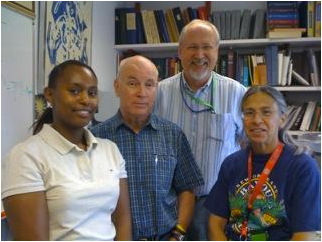
NICHD Biomedical Mass Spectrometry Facility Staff
Left to right: E. Ogbonna, A. Yergey, P. Backlund, N. Vieira
The facility's staff are available for consultation to everyone within the Institute, provided that existing resources are distributed equally among investigators requesting our services. Except for the ABI Voyager MALDI TOF (MALDI: matrix-assisted laser desorption/ionization; TOF: time-of-flight) instrument, none of the equipment is available for general use. The philosophy of the Facility is to ensure that its instruments record only reliable, high-quality data and that its clients receive only statistically meaningful analyses. For every new project, staff members meet with the Principal Investigator (PI) and the postdoctoral scientists involved in the study to discuss details of the experimental design. The discussion focuses on the project's background and analytical goals in order to determine the most appropriate techniques for sample preparation before delivery of samples to the laboratory. For protein characterizations, we digest the proteins in situ, typically gel-separated by the investigator, then isolate peptides for mass-spectrometric analysis. We subject the peptides to analysis by fragmenting them in a second stage of mass spectrometer operation and then use the fragmentation spectra as input for database search engines to determine the protein(s) present in the original sample. Upon completion of an analysis, we provide the investigators with the results and review the findings. Long-term user feedback indicates that our high level of interaction with researchers has greatly improved the quality and utility of results of each project undertaken.
Instrumentation
The facility currently has four mass spectrometers in use for specific areas of analysis.
- ABI 4800 MALDI TOF/TOF. This state-of-the-art high-performance instrument can be operated in both positive- and negative-ion modes. We typically use it for peptide mixtures without chromatographic separation. Additional uses include accurate determination of peptide masses in complex mixtures and the determination of sequences of unusual peptides through de novo sequencing techniques.
- Thermo Finnigan LCQ Deca
- LCQ DecaXP. Both the Thermo Finnigan LCQ Deca and the LCQ DecaXP employ liquid chromatography and electrospray ionization to separate and ionize peptides that are then fragmented. At present the DecaXP instrument is dedicated to a project on cardiolipin quantification in serum.
- ABI Voyager MALDI TOF. This instrument is used for analysis of higher molecular-weight species—equal to or less than 125 kDa—and to verify the molecular weight of intact proteins. The instrument is also available for general use after a potential user has undergone appropriate training.
Facility usage
The Mass Spectrometry Facility currently serves 15 sections within the institute, representing about 40 projects, as well as four PIs of sister institutes on the NIH campus.
Major projects
Brain Proteomics in Niemann-Pick Disease Type C (NPC). We have characterized cerebellar tissue from five-week-old littermate control (n=7) and Npc1-mutant female mice (Npc1m1N, n=7). After statistical analysis of 871 protein spots on quadruplicate gels, a total of 52 protein spots were found to be significantly increased (R≥1.20, p<0.05) and 12 protein spots significantly decreased (R≤0.83, p<0.05) in Npc1−/− cerebellar tissue. Differentially expressed protein spots were isolated from paired mutant and control preparative gels; using mass-spectrometric techniques, we have, to date, candidate identifications on 20 pairs of protein spots. Although validation is pending, one of the identified proteins is transthyretin. Although this protein identification was made in mouse brain, transthyretin is an intriguing potential biomarker in human CSF, one reason being that decreased levels of cerebral spinal fluid transthyretin correlate with disease severity in Alzheimer's disease. These studies have now been extended to one- and three-week-old littermates and a total of 200 additional differentially expressed gel spots.
Cerebral Spinal Fluid Profiling (CSF) in NPC. Initial experiments demonstrated that we can detect significant differences between cerebral spinal fluid from NPC patients and adult controls, using our novel analytical approach to compare protein profiles. CSF samples were collected from 37 well-characterized NPC patients. Currently, the collection for NPC patients includes longitudinal samples collected over 2.5 years and five patients pre/post miglustat treatment. In addition, collection of pediatric CSF samples is in progress at Children's National Medical Center. Our analytical approach accounts for systemic sources of variance, employs an analysis of variance (ANOVA) approach to the sets of MALDI-TOF spectra, and then applies Principal Component Analysis (PCA) to the data arrays generated by ANOVA. The results of ANOVA-PCA analyses will be correlated with patient disease status using a neurological severity scale that can be used to define disease status and progression in NPC patients, as developed by the Section on Molecular Dysmorphology, NICHD. The extensive differentially observed profile spectra are being further analyzed using mathematical tools of Fuzzy multivariate rule-building expert systems—minimal neural networks (FuRES)—to elucidate potential specific biomarkers. Initial results of this analysis show the presence of at least 10 specific peptide masses that are candidate biomarkers. We will employ chromatographic separation of the CSF followed by mass spectrometry to identify specific peptides in the CSF suggested by the 95% Confidence Intervals of the FuRES analysis.
Lipid Quantification in Serum. We have continued developing methodology to quantify cardiolipins in human serum by mass spectrometry. This effort is conducted in association with a clinical study under way in the Institute to evaluate the effects of antibiotic treatment of pregnant women colonized with the Group B streptococcal (GBS) organism. The hypothesis of the study is that the typical peri-natal penicillin treatment gives rise to a large increase of circulating cardiolipins in the infant, which then leads to respiratory distress. It has been demonstrated in other studies in newborn sheep that the GBS organisms secrete a specific cell-wall membrane cardiolipin upon penicillin treatment and that this substance causes respiratory distress at levels corresponding to the injection of about 100 pmole/mL in serum; this concentration falls rapidly with a half life of a few minutes. It is not known whether the respiratory distress observed in a fraction of infants born to GBS-colonized mothers is a result of a similar effect, or perhaps of a related effect caused by a release of endogenous cardiolipins stimulated by bacterial death. The analytical approach involves the addition of an internal standard to a serum sample, extractions by a combination of liquid-liquid and solid phase, followed by an LC-MS analysis that incorporates an extraction/recovery standard to monitor system quality control. We showed that cardiolipin can be extracted from serum with approximately 90% efficiency. We further showed that normal adult levels of (18:2)4 cardiolipins are present in serum at levels of less than 10 fmole/uL, approximately 1000-fold lower than found by earlier, less accurate measurements. With this base-line information, we are seeking to establish the serum levels in cord blood of mothers known not to be colonized by GBS and then to determine levels in infected individuals.
Community outreach
The Mass Spectrometry Facility is committed to promoting mass-spectrometric aspects of proteomics and other mass-spectrometric analyses in NICHD's Division of Intramural Research. We make serious efforts to educate investigators on the benefits and pitfalls of the techniques used in the Facility. In particular, we provide coaching on the principles of appropriate methods for the isolation and staining of gels and support an NIH-wide seminar series featuring internationally known experts in proteomics. In parallel, the staff of the Facility have developed collaborations with other Institutes to promote exchange of information and to bring new mass-spectrometric techniques to NICHD.
Additional Funding
- Bench to Bedside (2010): Development of combination therapy for Niemann-Pick Disease, type C
Publications
- Jiang X-S, Wassif CA, Backlund PS, Song L, Holtzclaw LA, Zheng L, Yergey AL, Porter FD. Activation of Rho GTPases in Smith-Lemli-Opitz syndrome: pathophysiological and Clinical Implications. Hum Mol Gen. 2010 19:1347-1357.
- Jiang X-S, Backlund PS, Wassif CA, Yergey AL, Porter FD. Quantitative proteomic analysis of inborn errors of cholesterol synthesis: identification of altered metabolic pathways in DHCR7 and SC5D deficiency. Mol Cell Prot. 2010 9:1461-1475.
Collaborators
- Paul Blank, PhD, Program on Physical Biology, NICHD, Bethesda, MD
- Jonathan Epstein, MS, Unit on Biologic Computation, OSD, NICHD, Bethesda, MD
- Peter Harrington, PhD, Ohio University, Athens, OH
- Matthew Olson, MD, The Johns Hopkins University Medical School, Baltimore, MD
- Forbes Porter, MD, PhD, Program on Developmental Endocrinology and Genetics, NICHD, Bethesda, MD
- John Robbins, MD, Program on Developmental and Molecular Immunity, NICHD, Bethesda, MD
- Dan Sackett, PhD, Program on Physical Biology, NICHD, Bethesda, MD
Contact
For more information, email aly@helix.nih.gov.