

You are here: Home > Mass Spectrometry Core Facility
NICHD Biomedical Mass Spectrometry Facility

- Alfred L. Yergey, PhD, Principal Investigator, Director (Emeritus)
- Peter S. Backlund, PhD, Staff Scientist, Acting Director
- Nancy E. Vieira, MS, Senior Research Assistant
- Stephanie Cologna, PhD, Intramural Research Training Award Fellow
- Christopher A. Crutchfield, PhD, Intramural Research Training Award Fellow
- Elizabeth Kahuno, BS, Contractor
The NICHD Biomedical Mass Spectrometry Core Facility was created under the auspices of the Office of the Scientific Director to provide high-end mass-spectrometric services to scientists within the NICHD Intramural Research Program (IRP). Particular focus has been in the areas of proteomics, biomarker discovery, protein characterization, and detection of post-translational modifications. The Facility also performs quantitative analyses of small bio-molecules, including lipids and steroids. The Facility is located in the 9D corridor of Building 10 on the NIH campus.
Mode of operation
Click image to enlarge.
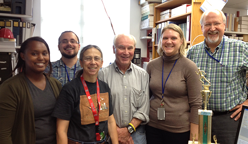
BMS Facility Staff
(L to R) E. Kahuno, C. Crutchfield, N. Vieira, A. Yergey, S. Cologna, and P. Backlund.
The Facility's staff are available for consultation with all labs within the Institute's IRP, provided that existing resources are distributed equally among investigators requesting our services. The philosophy of the Facility is to ensure that its instruments obtain only reliable, high-quality data and that its clients receive only statistically meaningful analyses. For each project, staff members meet with the Principal Investigator (PI) and other postdoctoral scientists involved in the study to discuss the experimental design. The discussion focuses on the project's background and analytical goals in order to determine the appropriate techniques for sample preparation and analysis, before delivery of samples to the Facility. For protein characterization, we analyze 1D- and 2D-PAGE–separated proteins (typically gel-separated by the investigator), as well as more complex mixtures from biological fluids. Depending on sample complexity, the peptides can be analyzed by either MALDI TOF-TOF or LC ESI-MS/MS methods. Upon completion of an analysis, we provide investigators with the results and review the findings. Long-term user feedback indicates that our high level of interaction with researchers greatly improves the quality and utility of results of each project undertaken.
Instrumentation
The facility currently has five mass spectrometers in use for specific areas of analysis.
- ABI 4800 MALDI TOF/TOF: The state-of-the-art high-performance instrument can be operated in both positive- and negative-ion modes and is used for peptide identification for peptide mixtures without chromatographic separation. Methodology is also available to perform off-line LC separation and sample spotting. Additional uses include relative peptide quantification for iTRAQ–labeled peptides, and sequence determination through de novo sequencing techniques for unusual peptides not present in gene-based protein databases.
- Agilent 6520 LC-ESI qTOF: The state-of-the art hybrid instrument produces high-resolution MS and MS/MS spectra for a variety of discovery projects. Current applications include the analysis of serum cardiolipins, protein MW characterization, and peptide identification.
- Agilent 6460 LC-ESI QqQ (Triple Quad): The state-of-the-art triple quad instrument is currently being used for small-molecule analysis and quantification, principally for urinary steroid profiling and the determination of glycolytic pathway metabolites.
- Thermo-Finnigan LCQ Deca: This lower-resolution instrument is used exclusively for peptide characterization by nanoLC–electrospray ionization and fragmentation. It yields data that are complimentary to those produced by the MALDI TOF/TOF.
- ABI Voyager MALDI TOF: The instrument is used for the analysis of protein mixtures, and to verify molecular weights of intact proteins. The instrument is also available for general use after a potential user has undergone appropriate training.
Facility usage
The Mass Spectrometry Facility currently serves 15 sections within the institute, representing about 40 projects, as well as four PIs of sister institutes on the NIH campus.
Major projects
Proteomics of Mouse Models for Human Diseases: Proteomic studies were performed using mouse models in order to gain a further understanding of the pathophysiology for two human diseases: a) fatal, neurodegenerative Niemann-Pick disease-Type C1 (NPC1), and b) tumor aggressiveness of pheochromocytomas/paragangliomas. For each model system, proteins were separated by 2-dimentional gel electrophoresis (2D-GE) and quantified by the intensity of spot staining. Differentially expressed protein spots (> 1.5 fold change, p<0.05) were then excised and identified by mass spectrometric techniques.
We compared mouse cerebellum tissues from Npc1 knock-out with those from wild-type animals at 1, 3, and 5 weeks of age. Protein identities were obtained for 77 differentially expressed proteins. The proteins include those involved in glucose metabolism, detoxification/oxidative stress, and Alzheimer disease–related proteins. Members of the fatty acid binding protein family, FABP3, FABP5, and FABP7, were also found to have altered expression. With regard to the cerebral spinal fluid (CSF) from NPC1 patients, glutathione-S-transferase a, superoxide dismutase, and FABP3, were found to have altered expression compared with control CSF.
Comparing aggressive and non-aggressive mouse tumor cell lines from mouse PHEO cells, fifty-three differentially expressed proteins were observed and identified by mass spectrometry. Ingenuity pathway analysis indicated that the OXPHOS pathway was the pathway with the most significant changes, with four spots identified as subunits of OXPHOS complexes, including ubiquinol-cytochrome-c reductase protein 1, and ATP synthase β, γ, and δ subunits. Expression of the glycolysis-related protein lactate dehydrogenase B decreased in the aggressive tumor cell line, while expression of lactate dehydrogenase A and SOD2 increased. The later two proteins have been identified as promising therapeutic targets in other cancers.
Protein Profiling and Quantification in CSF: Initial experiments on protein profiling detected significant differences between proteins in CSF from NPC patients and adult controls using a novel analytical approach that we developed, by which MALDI spectra were analyzed by combining ANalysis Of Variance with Principal Component Analysis (ANOVA-PCA). We are now developing a method to compare relative levels of specific proteins in CSF from patients, using an 8-plex iTRAQ labeling process to mass-tag peptides generated from proteins present in each sample. The method will maintain individual patient information and permit comparison of results across multiple iTRAQ experiments. Initially, we are studying the Npc1 knock-out mouse model to determine the analytical variation of this method. Several proteins identified in our pilot study were also identified in our previous study of differentially expressed proteins in the cerebella of the mutant mice by 2D-GE. One example of a differentially expressed protein detected by both methods was ATP-synthase alpha, which showed a 2.6-fold decrease (p=0.002) by 2D-GE while initial iTRAQ experiments detected a 1.7-fold decrease (p=0.07). An additional protein that was found only in the iTRAQ experiment was Purkinje Cell Protein 4, which was down-regulated 1.9-fold (p=0.02) in the mutant mouse. Purkinje cells are known to be the first neuronal cell type to succumb in NPC1 disease. After completion of the variability study, this method will be used to profile protein changes in CSF from patients with NPC1.
Lipid Characterization and Quantification in Serum: We developed a methodology to extract and quantify cardiolipins in human serum. This effort is being carried out in support of a clinical study in the Institute to evaluate the effects of antibiotic treatment for pregnant women colonized with Group B streptococcal (GBS) organism on serum cardiolipins. The hypothesis of the study is that the typical peri-natal penicillin treatment gives rise to a large increase of circulating cardiolipins in the infant, which then leads to respiratory distress. The analytical approach involves addition of an internal standard to a serum sample, followed by liquid-liquid and solid-phase extractions. The cardiolipins are then detected and quantified by LC-MS analysis. Using this method, we analyzed cardiolipins in normal adult serum and determined concentrations of (18:2)-4 cardiolipin in a range of 3–10 nM, approximately 1000-fold lower than reported by earlier, less accurate measurements. In addition, cardiolipins with other fatty acid moieties have been detected. The method is currently being applied to quantify cardiolipins in maternal serum, cord blood, and infant serum from the clinical study.
New Approach to Mass Spectrometry-Based Protein Identification: Current approaches to protein identification rely on database matching of fragmentation spectra, while ignoring (in a scoring sense) the mass accuracy of the precursor ions. We developed a method that uses targeted peptide mass fingerprinting to confirm MS/MS database search identifications. The distribution of mass errors of matches in the first order spectrum is used to develop a probability model that is independent from MS/MS database searches, employing Bayesian Inference and the mass errors in the first-order spectra to assign peptide scores. Products of the peptide probabilities yield the final protein scores. These true protein scores are then assigned a probability of being correct by applying Extreme Value Theory to a set of randomly selected decoy proteins that have approximately the same mass as the true proteins but have scrambled sequences. The method was tested on a mixture of protein standards, and results demonstrated both very high confidence levels for proteins identified by MS/MS fragmentation and validated several low scoring "one-hit wonders" found by MS/MS fragmentation.
Mass Spectrometry–Based Profiling of Urinary Steroids: Current approaches to the analysis of urinary steroids typically employ either immunoassay or mass spectrometry–based technologies. Immunoassay-based methods often lack specificity owing to cross-reactivity with other steroids, and targeted LC-MS/MS is limited to the analysis of pre-determined analytes. We developed a new LC-MS/MS approach to urinary steroid profiling that enables us to detect the steroids that have truly changed in a patient cohort without knowing their identity beforehand (i.e., untargeted metabolomics of steroids). In addition, we utilized a product ion spectrum database of known steroids to improve our ability to identify novel steroids. We tested the methods in a pilot project to investigate urinary steroids of patients diagnosed with polycystic ovarian syndrome (PCOS). Initially, the studies detected elevated levels of an unknown compound consistent with an androgenic steroid in PCOS patients. We were then able to identify the unknown as a mixture of androsterone-sulfate and etiocholanolone-sulfate. Two-dimensional liquid chromatography, using C18 in the first dimension and Porous Graphitic Carbon in the second, was required to separate the diastereomeric pair.
Community outreach
The Mass Spectrometry Facility is committed to promoting mass-spectrometric aspects of proteomics and other mass-spectrometric analyses in NICHD's Division of Intramural Research. We make serious efforts to educate investigators on the benefits and pitfalls of the techniques used in the Facility. In particular, we provide coaching on the principles of appropriate methods for sample isolation and staining of gels. We also support an NIH–wide seminar series featuring internationally known experts in proteomics. In parallel, the staff of the Facility have developed collaborations with other Institutes to promote exchange of information and to bring new mass-spectrometric techniques to NICHD. In addition, Peter Backlund is the coordinator of the NIH Mass Spectrometry Interest Group.
Publications
- Fliedner SM, Kaludercic N, Jiang XS, Hansikova H, Hajkova Z, Sladkova J, Limpuangthip A, Backlund PS, Wesley R, Martiniova L, Jochmanova I, Lendvai NK, Breza J, Yergey AL, Paolocci N, Tischler AS, Zeman J, Porter FD, Lehnert H, Pacak K. Warburg effect's manifestation in aggressive pheochrocytomas and paragangliomas: insights from a mouse cell model applied to human tumor tissue. PLoS One 2012;7:e40949.
- Neunuebel MR, Chen Y, Gaspar AH, Backlund P, Yergey A, Machner MP. De-AMPylation of the small GTPase Rab1 by the pathogen Legionella pneumophila. Science 2011;333:453-456.
- Olson MT, Epstein JA, Sackett DL, Yergey AL. Production of reliable MALDI spectra with quality threshold clustering of replicates. J Am Soc Mass Spect 2011;11:969-975.
Collaborators
- Paul Blank, PhD, Program in Physical Biology, NICHD, Bethesda, MD
- Jonathan Epstein, MS, Molecular Genomics Laboratory Core Facility, NICHD, Bethesda, MD
- Peter Harrington, PhD, Ohio University, Athens, OH
- Rachel O. Loo, PhD, Molecular Biology Institute, UCLA, Los Angeles, CA
- Mattias Machner, PhD, Cell Biology and Metabolism Program, NICHD, Bethesda, MD
- Matthew Olson, MD, The Johns Hopkins University Medical School, Baltimore, MD
- Forbes Porter, MD, PhD, Program in Developmental Endocrinology and Genetics, NICHD, Bethesda, MD
- John Robbins, MD, Program in Developmental and Molecular Immunity, NICHD, Bethesda, MD
- Dan Sackett, PhD, Program in Physical Biology, NICHD, Bethesda, MD
- Constantine A. Stratakis, MD, DSc, Program in Developmental Endocrinology and Genetics, NICHD, Bethesda, MD
Contact
For more information, email backlunp@mail.nih.gov.