You are here: Home > Section on Physical Biochemistry
Extracellular Matrix Disorders: Molecular Mechanisms and Treatment Targets

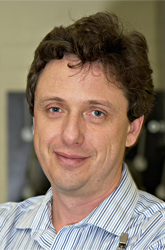
- Sergey Leikin, PhD, Head, Section on Physical Biochemistry
- Edward L. Mertz, PhD, Staff Scientist
- Elena N. Makareeva, PhD, Postdoctoral Fellow
- Lynn Felts, BS, Predoctoral Fellow (Graduate Student)
- Aaron Konopko, BA, Postbaccalaureate Fellow
The extracellular matrix (ECM) is involved in a wide variety of disorders, ranging from rare genetic abnormalities of skeletal development (skeletal dysplasias) to such common ailments as osteoporosis, fibrosis, and cancer. Our interest in ECM biology began with studies on basic principles relating the helical structure of collagen and DNA to their interactions and biological function. We continue to study DNA–DNA interactions, particularly sequence recognition in homologous recombination. However, the focus of our research gradually shifted to collagens, which are the most abundant ECM molecules, and then to molecular mechanisms underlying disorders involving collagens and other ECM proteins and proteoglycans. We investigate how abnormalities in these molecules contribute to cancer, fibrosis, osteogenesis imperfecta, Ehlers-Danlos syndrome, chondrodysplasias, osteoporosis, and other diseases. Together with other NICHD and extramural clinical scientists, we strive to improve our knowledge of the molecular mechanisms underlying these diseases. We hope to use the knowledge gained through these studies for diagnostics, characterization, and treatment, bringing our expertise in physical biochemistry and theory to clinical research and practice.
Procollagen folding and its role in bone disorders
Collagens are triple-helical proteins forming structural scaffolds that hold together bone, cartilage, skin, and other tissues. In bone, the proteins are produced by osteoblasts, the cells responsible for synthesis and mineralization of new bone material. By far the most abundant proteins in all vertebrates, they account for up to 30% of all proteins synthesized by osteoblasts. Yet, folding of their procollagen precursors within the endoplasmic reticulum (ER) presents an extraordinary challenge for cells. In particular, we discovered that the equilibrium state of type I procollagen in the absence of chaperones is a random coil. Folding of the human procollagen triple helix in an ER–like environment is favorable below 35°C but not at normal body temperature. At body temperature, natively folded collagen molecules become stable only after incorporation into fibers. Individual molecules, not incorporated into fibers denature within several hours and become susceptible to rapid proteolytic degradation. To overcome such intrinsic instability, cells must use specialized chaperones to fold the triple helix. It is not completely clear which chaperones are involved in this process, how they function, and how cells recognize properly folded procollagen and handle it when misfolded. Because of massive procollagen synthesis, osteoblast malfunction caused by procollagen misfolding might be an important factor in bone pathologies. For instance, osteogenesis imperfecta appears to be primarily a procollagen misfolding disease. Moreover, inability of aging osteoblasts to handle normal procollagen folding load might also contribute to common osteoporosis. Understanding procollagen folding and cellular response to its misfolding is therefore important both from fundamental and practical perspectives (1).
The most common mutations affecting procollagen folding are Gly substitutions in the obligatory (Gly-X-Y)n sequence of the triple helix. Such substitutions in type I collagen are, for example, responsible for approximately 80 % of severe osteogenesis imperfecta (OI) cases. The substitutions' effects on procollagen folding are determined by the location and identity of the substituting residue. Our studies of collagens from OI patients with over 50 different Gly substitutions revealed several structural regions within the triple helix where mutations might be responsible for distinct OI phenotypes. In particular, the first 85–90 amino acids at the N-terminal end of the triple helix form an "N-anchor" domain with higher-than-average triple helix stability. Gly substitutions within this region disrupt the whole N-anchor, preventing normal cleavage of the adjacent N-propeptide. Incorporation of collagen with uncleaved N-propeptides into fibrils leads to hyperextensibility and joint laxity more characteristic of the Ehlers-Danlos syndrome (EDS). Gly substitutions within an alpha1(I) chain region surrounding the collagenase cleavage site might be lethal because the sequence of this region prevents efficient renucleation of the normal C-to-N-terminus helix folding once the mutation is encountered.
At present, we are focusing on understanding how cells respond to misfolding of procollagen with different Gly substitutions. We hypothesize that unconventional procollagen triple helix folding requirements lead to unconventional ER stress response and to misfolding (1). Our studies indicate that misfolded procollagen molecules are preferentially retained within the ER and targeted for intracellular degradation without activation of the conventional unfolded protein response (UPR). We observe accumulation of mutant molecules within the ER, but no upregulation of UPR chaperones (like BiP) or UPR signaling pathways (like XBP1 splicing). Instead, our preliminary data support dramatic upregulation of HSP47, activation of autophagy, and involvement of NF-kB signaling reminiscent of a so-called ER overload response typical of serpinopathies. Much work remains to be done to understand the underlying signaling pathways and identify potential targets for therapeutic intervention.
Translational studies of patients with novel or unusual OI mutations
Deficiencies in ER chaperones involved in procollagen folding form the second largest group of mutations causing severe OI. These proteins include but may not be limited to HSP47, FKBP65, cartilage-associated protein (CRTAP), prolyl-3-hydrohylase (P3H1), and cyclophilin B (CYPB). In collaboration with scientists from the Bone and Extracellular Matrix Branch of NICHD, we characterized effects of CRTAP/P3H1/CYPB complex as well as FKBP65 deficiencies on procollagen folding and extracellular matrix deposition (1–3). However, molecular mechanisms underlying these effects and resulting cellular responses remain largely unclear.
A particularly puzzling form of OI is associated with deficiencies in the pigment epithelium–derived factor (PEDF). The protein was initially described as an anti-angiogenesis factor, but the loss of function of both PEDF alleles leads to severe, progressively deforming OI (type VI) rather than out-of-control angiogenesis. Our collaborative study with the clinical researcher Frank Rauch revealed no abnormalities in procollagen folding or secretion by fibroblasts from type VI OI patients. At the same time, our collaborative study with Patricia Becerra showed that PEDF is the most potent of all known inhibitors of collagen fiber formation. At normal serum concentration (about 100 nM), PEDF slows down collagen fiber formation more than ten-fold, suggesting that fibrillogenesis control might be one of its physiological functions. However, a recently reported type VI OI patient without PEDF mutations and thus with normal PEDF concentration in the serum but deficient in PEDF synthesis in osteoblasts suggests that PEDF might affect osteoblast differentiation and/or function from inside. We are pursuing this idea with PEDF knockdown experiments in cultures of primary human bone marrow stromal cells as well as immortalized human and mouse pre-osteoblasts.
Murine models of bone disorders
Mouse models offer unique opportunities for more systematic studies of molecular mechanisms underlying bone disorders. Our studies of mouse OI models with knock-in alpha1(I)-G349C and alpha2(I)-G610C substitutions revealed significant ER stress and resulting osteoblast malfunction caused by misfolding of mutant procollagen. We are focusing on understanding molecular mechanisms of ER–stress response to this misfolding and their contribution to bone pathology. One idea is that osteoblast function in these animals might be improved by reducing procollagen misfolding load, achieved by stimulating the autophagy responsible for eliminating misfolded molecules from the ER. We are testing the simplest practical approach to in vivo autophagy enhancement in G610C animals based on a reduced protein diet supplemented with methionine and aromatic amino acids essential for bone. In addition to providing information on the suspected role of autophagy in ER–stress response to procollagen misfolding, this approach might offer some relief for OI patients and provide an additional tool for the management of age-related osteoporosis. If successful, its simplicity and safety would enable quick clinical trials and implementation.
In the past several years, we also collaborated with Program in Developmental Endocrinology and Genetics (PDGEN) scientists at NICHD to investigate bone tumors caused by defects in protein kinase A (PKA), a crucial enzyme in the cAMP signaling pathway. We found that knockouts of various PKA subunits cause not only abnormal organization and mineralization of bone matrix but also novel bone structures that had not been reported before. For instance, we observed free-standing cylindrical bone spicules with osteon-like organization of lamellae and osteocytes but inverted mineralization pattern, highly mineralized central core, and decreasing mineralization away from the central core. Currently, we are assisting PDGEN scientists in characterizing abnormal osteoblast maturation, the role of abnormal inflammatory response, and effects of anti-inflammatory drug treatments in these animals. Improved understanding of bone tumors caused by PKA deficiencies may not only clarify the role of cAMP signaling but also suggest new approaches to therapeutic manipulation of bone formation in skeletal dysplasias.
Collagen matrix pathology in cancer and fibrosis
An important advance of the past several years was the characterization of a collagenase-resistant, homotrimeric isoform of type I collagen and its potential role in cancer, fibrosis, and other disorders. The normal isoform of type I collagen is a heterotrimer of two alpha1(I) chains and one alpha2(I) chain. Homotrimers of three alpha1(I) chains are produced in some fetal tissues, carcinomas, fibrotic tissues, as well as in rare forms of OI and EDS associated with alpha2(I) chain deficiency. We found the homotrimers to be at least 5–10 times more resistant to cleavage by all mammalian collagenases than the heterotrimers and we determined the molecular mechanism of this resistance. Our studies suggested that cancer cells might utilize this collagen isoform for building collagenase-resistant tracks, supporting invasion through stroma of lower resistance. Homotrimer production by bone marrow stromal cells as well as embryonic and immature osteoblasts might also contribute, for example, to increased severity of alpha1(I) mutations in OI and bone tumors in PKA deficiencies. Currently, we are investigating the regulation of the homotrimer synthesis by various cells and developing approaches to selective targeting of homotrtimers.
Our studies on type I collagen homotrimers in various human tumors and fibrotic lesions unexpectedly uncovered another potentially important collagen pathology. In collaboration with scientists from the Program in Reproductive and Adult Endocrinology, NICHD, we discovered abnormally high content of the alpha3(V) chain of type V collagen in more aggressive forms of pheochromocytomas and paragangliomas. Given the suspected role of collagen molecules containing this chain in glucose metabolism and abnormalities of glucose metabolism in these tumors, this finding opens a promising research opportunity. We hope that a better research funding environment will enable us to pursue this opportunity in the future.
High-definition infrared and Raman micro-spectroscopy of tissues
Label-free micro-spectroscopic infrared and Raman imaging of tissues and cell cultures provides important information about chemical composition, organization, and biological reactions inaccessible by more traditional histology. However, application of the technologies had been restricted by the limited accuracy of spectra from hydrated specimens under physiological conditions. We resolved this problem by thermo-mechanical stabilization of specimen chambers, reducing path-of-light instabilities and improving spectral reproducibility by over two orders of magnitude. This high-definition (HD) technology was crucial for our studies on bone structure and mineralization in mouse models of bone disorders described above. It was essential for analysis of abnormal collagen matrix deposition by FKBP65–deficient cells (3). It also enabled us to assist NIBIB scientists in characterizing a functionalized carbon nanotube approach to delivery of anticancer agents into cells that overexpress hylauronate receptors (4).
The power of this technology is best illustrated by our studies on cartilage pathology in a mouse model of dyastrophic dysplasia (DTD) caused by mutations in the SLC26A2 sulfate/chloride antiporter that affect sulfation of cartilage proteoglycans (5). In DTD mice, proteoglycan sulfation is only slightly reduced at birth and normalizes with age, but articular cartilage progressively degrades with age, and bones develop abnormally. Our HD–infrared imaging revealed strong chondroitin undersulfation within narrow regions of the growth plate and periarticular region. This undersulfation disrupted, e.g., formation of a thin layer of well oriented collagen fibers at the articular surface in newborn mice. The disruption likely leads to insufficient cartilage protection from mechanical damage and synovial enzymes, potentially explaining the observed progressive cartilage degradation with animal age despite normalization of chondroitin sulfation. By correlating HD–infrared and micro-autoradiographic imaging, we found that accelerated chondroitin synthesis in cartilage regions crucial for bone growth may cause a significant deficit of intracellular sulfate in DTD. Such a deficit may also contribute to the cartilage and bone development pathology by affecting sulfation of heparan and other glycosaminoglycans essential for cell signaling.
Theory and measurements of sequence-dependent DNA–DNA interactions
Given the fundamental importance and practical applications of DNA–DNA interactions, we continue to invest a small fraction of our resources into theoretical studies on such interactions and analysis of the corresponding experiments reported in the literature. Interactions between double-stranded (duplex) DNA molecules are usually presumed to be independent of the DNA sequence because the nucleotides are buried inside the double helix and shielded by the highly charged sugar-phosphate backbone. However, our theory of electrostatic interactions between molecules with helical patterns of surface charges revealed significant effects of the sequence-dependent double helix structure on DNA–DNA interactions. These effects arise from preferential juxtaposition of the negatively charged sugar phosphate backbone with positively charged counterions bound in grooves on the opposing molecule. The theory explained the observed counter-ion specificity of DNA condensation, changes in DNA structure upon aggregation, multiple packing arrangements and measured osmotic pressures in DNA aggregates, as well as observed positions and shapes of peaks in x-ray diffraction patterns from hydrated DNA fibers. Our most recent study on interactions between oligonucleotide nucleic acids suggested an explanation for puzzling observations of double-stranded (ds) RNA resistance to condensation by cobalt hexamine, which condenses ds–DNA. It also indicated why triple-stranded DNA oligonucleotides are condensed by divalent alkaline earth metal ions, which do not condense ds–DNA. Direct recognition of sequence homology between 100 base-pair (bp) or longer sequences and pairing of intact double helices without unwinding predicted by our theory may have particularly important biological implications. In vitro, this prediction was supported by our experiments done in collaboration with scientists from the Imperial College, London, UK, as well as by observations reported by others. We hope that a better research funding environment in the future will enable us to set up an experimental group at NICHD dedicated to investigating such homologous pairing of DNA double helices in live cells.
Publications
- Makareeva E, Aviles NA, Leikin S. Chaperoning osteogenesis: new protein-folding disease paradigms. Trends Cell Biol 2011;21:168-176.
- Valli M, Barnes AM, Gallanti A, Cabral WA, Viglio S, Weis MA, Makareeva E, Eyre D, Leikin S, Antoniazzi F, Marini JC, Mottes M. Deficiency of CRTAP in non-lethal recessive osteogenesis imperfecta reduces collagen deposition into matrix. Clin Genet 2012;82:453-459.
- Barnes AM, Cabral WA, Weis M, Makareeva E, Mertz EL, Leikin S, Eyre D, Trujillo C, Marini JC. Absence of FKBP10 in recessive type XI osteogenesis imperfecta leads to diminished collagen cross-linking and reduced collagen deposition in extracellular matrix. Hum Mutat 2012;33:1589-1598.
- Swierczewska M, Choi KY, Mertz EL, Huang X, Zhang F, Zhu L, Yoon HY, Park JH, Bhirde A, Lee S, Chen X. A facile, one-step nanocarbon functionalization for biomedical applications. Nano Lett 2012;12:3613-3620.
- Mertz EL, Facchini M, Pham AT, Gualeni B, De Leonardis F, Rossi A, Forlino A. Matrix disruptions, growth, and degradation of cartilage with impaired sulfation. J Biol Chem 2012;287:22030-22042.
Collaborators
- S. Patricia Becerra, PhD, Laboratory of Retinal Cell and Molecular Biology, NEI, Bethesda, MD
- Peter H. Byers, MD, University of Washington, Seattle, WA
- Antonella Forlino, PhD, Università degli Studi di Pavia, Pavia, Italy
- Alexei A. Kornyshev, PhD, Imperial College, University of London, London, UK
- Joan C. Marini, MD, PhD, Bone and Extracellular Matrix Branch, NICHD, Bethesda, MD
- Hideaki Nagase, PhD, Imperial College, University of London, London, UK
- Karel Pacak, MD, PhD, DSci, Program in Reproductive and Adult Endocrinology, NICHD, Bethesda, MD
- Charlotte L. Phillips, PhD, University of Missouri, Columbia, MO
- Frank Rauch, MD, Shriners Hospital for Children, Montreal, Canada
- Antonio Rossi, PhD, Università degli Studi di Pavia, Pavia, Italy
- Dan L. Sackett, PhD, Program in Physical Biology, NICHD, Bethesda, MD
- Constantine A. Stratakis, MD, DSci, Program in Developmental Endocrinology and Genetics, NICHD, Bethesda, MD
Contact
For more information, email leikins@mail.nih.gov or visit physbiochem.nichd.nih.gov.

