

You are here: Home > Laboratory on the Mechanism and Regulation of Protein Synthesis
The Molecular Mechanics of Eukaryotic Translation Initiation
- Jon Lorsch, PhD, Head, Laboratory on the Mechanism and Regulation of Protein Synthesis
- Jagpreet Nanda, PhD, Staff Scientist
- Sarah Walker, PhD, Research Fellow
- Fujun Zhou, PhD, Research Fellow
- Colin Aitken, PhD, Postdoctoral Intramural Research Training Awardee
- Shardul Kulkarni, PhD, Postdoctoral Intramural Research Training Awardee
- Antonio Munoz, BA, Predoctoral Intramural Research Training Awardee
- Paul Yourik, BA, Predoctoral Intramural Research Training Awardee
The goal of our research group is to elucidate the molecular mechanisms underlying the initiation phase of protein synthesis in eukaryotic organisms. We use the yeast Saccharomyces cerevisiae as a model system and employ a range of approaches—from genetics to biochemistry to structural biology—in collaboration with Alan Hinnebusch's and Tom Dever’s labs at NICHD and several other research groups around the world.
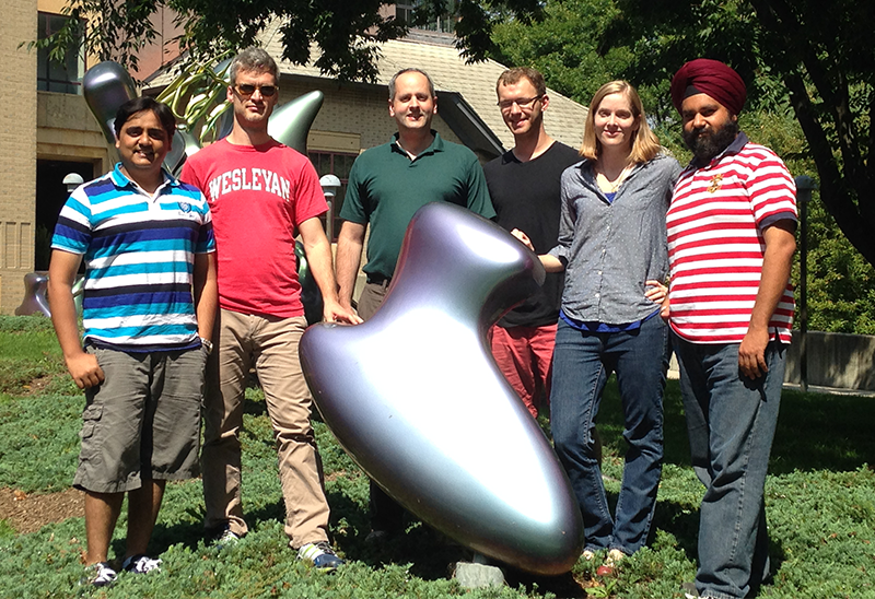
Lorsch Lab at Cold Spring Harbor Translational Control Meeting
Eukaryotic translation initiation is a key control point in the regulation of gene expression. It begins when an initiator methionyl tRNA (Met-tRNAi) is loaded onto the small (40S) ribosomal subunit. Met-tRNAi binds to the 40S subunit as a ternary complex (TC) with the GTP–bound form of the initiation factor eIF2. Three other factors, eIF1, eIF1A, and eIF3, also bind to the 40S subunit and promote the loading of the TC. The resulting 43S pre-initiation complex (PIC) is then loaded onto the 5′-end of an mRNA with the aid of eIF3 and the eIF4 group of factors: the RNA helicase eIF4A; the 5′-7-methylguanosine cap-binding protein eIF4E; the scaffolding protein eIF4G; and the 40S subunit– and RNA–binding protein eIF4B. Both eIF4A and eIF4E bind to eIF4G and form the eIF4F complex. Once loaded onto the mRNA, the 43S PIC is thought to scan along the mRNA in search of an AUG start codon. The process is ATP–dependent and likely requires multiple RNA helicases, including the DEAD–box protein Ded1p. Recognition of the start site begins with base pairing between the anticodon of tRNAi and the AUG codon. This base pairing then triggers downstream events that commit the PIC to continuing initiation from that point on the mRNA. These events include ejection of eIF1 from its binding site on the 40S subunit, movement of the C-terminal tail (CTT) of eIF1A, and release of phosphate from eIF2, which converts eIF2 to its GDP–bound state. In addition, the initiator tRNA moves from a position that is not fully engaged in the ribosomal P site [termed P(OUT)] to one that is [P(IN)], and the PIC as a whole converts from an open conformation that is conducive to scanning to a closed one that is not. At this stage, eIF2•GDP dissociates from the PIC, and eIF1A and a second GTPase factor, eIF5B, coordinate joining of the large ribosomal subunit to form the 80S initiation complex. In a process that appears to result in conformational reorganization of the complex, eIF5B hydrolyzes GTP and then dissociates along with eIF1A.
Advances in understanding the mechanism of start codon recognition
In collaboration with Alan Hinnebusch’s lab, we probed the functions of conserved identity element bases in the initiator tRNA (1). Our data indicate that each region of the tRNA plays important roles in start codon recognition. Changing a conserved G:C base pair in the anticodon stem to another base pair decreases the fidelity of start codon recognition (Sui− phenotype), whereas disrupting the base pair increases fidelity (Ssu− phenotype). The latter effect is suppressed by substitutions in a conserved base in the T-loop, indicating long-range communication within the tRNA body. These in vivo effects are mirrored by effects observed in vitro on the stability of TC binding to the 43S PIC, suggesting that the substitutions alter the relative stabilities of the P(OUT) and P(IN) states of the complex. Similarly, changes in the acceptor stem base pair C3:G70 reduce the fidelity of start codon recognition in vivo and perturb TC binding to the PIC in vitro. Overall, our results indicate that, over its entire length, the initiator tRNA structure is finely tuned to set the energetic bars for undergoing the conformational transitions within the PIC that are required for accurate start codon recognition.
We also probed the mechanism of action of eIF5, which is the GTPase–activating protein for eIF2. In addition to this function, eIF5 has also been shown to play an important role in start codon recognition. In collaboration with Alan Hinnebusch’s lab, we showed that the G31R mutation in eIF5, which reduces the fidelity of start codon recognition in vivo by specifically increasing utilization of UUG codons, increases the rate of release of inorganic phosphate from eIF2 in PICs at UUG codons and decreases it at AUG codons (2). The effect is consistent with our previous work, which indicated that the mutation stabilizes the closed state of the PIC at UUG codons and destabilized it at AUG codons. A suppressor of the G31R mutation, G62S, reverses both of these effects and restores a more normal AUG/UUG initiation ratio in vivo. A second suppressor mutation in eIF5, M18V, functions in a different manner by diminishing the ability of the factor to activate GTP hydrolysis by eIF2 without affecting the ability of PICs to convert from the open to closed states. Our data support the notion that eIF5 plays several key roles in start codon recognition: activating GTP hydrolysis, controlling phosphate release, and modulating the conformational state of the PIC.
In collaboration with Venki Ramakrishnan’s lab, we determined the structures of intermediate PICs at 4 Å resolution using cryo-electron microscopy (3). The structures shed considerable light on events that take place in the PIC in response to start codon recognition.
Publications
- Dong J, Munoz A, Kolitz SE, Saini AK, Chiu WL, Rahman H, Lorsch JR, Hinnebusch AG. Conserved residues in yeast initiator tRNA calibrate initiation accuracy by regulating preinitiation complex stability at the start codon. Genes Dev 2014;28:502-520.
- Saini AK, Nanda JS, Martin-Marcos P, Dong J, Zhang F, Bhardwaj M, Lorsch JR, Hinnebusch AG. Eukaryotic translation initiation factor eIF5 promotes the accuracy of start codon recognition by regulating Pi release and conformational transitions of the preinitiation complex. Nucleic Acids Res 2014;42:9623-9640.
- Hussain T, Llácer JL, Fernández IS, Munoz A, Martin-Marcos P, Savva CG, Lorsch JR, Hinnebusch AG, Ramakrishnan V. Structure of a eukaryotic translational initiation complex reveals key interactions in recognition of the start codon. Cell 2014;in press.
Collaborators
- Thomas Dever, PhD, Program in Cellular Regulation and Metabolism, NICHD, Bethesda, MD
- Alan Hinnebusch, PhD, Program in Cellular Regulation and Metabolism, NICHD, Bethesda, MD
- Venkatraman Ramakrishnan, PhD, MRC Laboratory of Molecular Biology, Cambridge, United Kingdom