

You are here: Home > Perinatology Research Branch
Mechanisms of Disease in Preterm Labor and Complications of Prematurity; Prenatal Diagnosis of Congenital Anomalies

- Roberto Romero, MD, Head, Program in Perinatal Research and Obstetrics
Preterm birth is the leading cause of perinatal morbidity and mortality worldwide. The Perinatology Research Branch proposed that preterm parturition is a syndrome caused by multiple pathologic processes, one of which is an untimely decline in progesterone action, manifested by "silent" shortening of the uterine cervix. Previous work conducted by our Program showed that asymptomatic women who have a cervical length less than 15mm in the second trimester of pregnancy have a 50% likelihood of delivering an early preterm neonate. However, prediction of preterm birth needs to be accompanied by a strategy to reduce the frequency of this complication. In a previous randomized clinical trial of vaginal progesterone vs. placebo in women with a short cervix, we reported that treatment was associated with a 45% reduction in the rate of preterm birth (less than 33 weeks of gestation), and a decrease in the rate of respiratory distress syndrome, the most common complication of prematurity. This year, we conducted studies to examine the relative merits of vaginal progesterone vs. cervical cerclage in patients with a short cervix and a prior history of preterm birth and found that medical treatment is as effective as the surgical approach. We also identified a novel pathologic finding associated with preterm labor without infection: chronic chorioamnionitis, which is likely attributable to maternal anti-fetal rejection, and we described a novel form of systemic fetal inflammation.
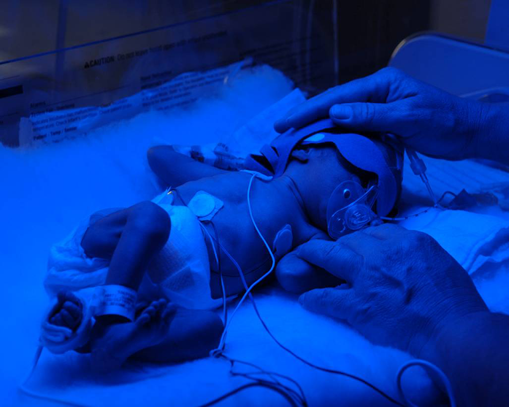
Click image to enlarge.
Figure 1. Preterm birth is the leading cause of perinatal mortality and morbidity worldwide and affects 12% of all pregnancies in the United States.
Other great obstetrical syndromes responsible for the high rate of infant mortality in the United States include fetal death and preeclampsia. This year, we reported that a blood test performed at the beginning of the third trimester can, with 80% sensitivity and 94% specificity, identify mothers who will have a stillbirth near term. In addition, we published findings that an anti-angiogenic profile is present in patients who have a maternal floor infarction, a condition associated with fetal growth restriction and fetal death.
Congenital anomalies continue to be a leading cause of perinatal mortality in the United States. Congenital heart disease is the leading organ-specific birth defect, as well as the leading cause of infant mortality from congenital malformations. More than half the infants affected with congenital heart disease are born to mothers without any previously known risk factors, which provides the impetus to perform a comprehensive screening examination of the fetal heart in all pregnancies. Yet, the prenatal diagnosis of congenital heart disease remains a challenge, as the sensitivity of ultrasound has ranged from 15–39%. The lack of prenatal identification of congenital heart defects can have adverse consequences for the neonate. Examination of the fetal heart is time-consuming, and expertise and skill are required. Therefore, the examination frequently does not include all the standard recommended cardiac views. Spatiotemporal image correlation (STIC) technology allows the acquisition of a volume dataset from the fetal heart, and displays a cine loop of a complete single cardiac cycle in motion. A growing body of evidence suggests that 4-D sonography with STIC facilitates examination of the fetal heart. However, extracting and displaying the recommended diagnostic planes from a volume dataset that can be dissected in many ways (i.e., planes) requires an in-depth knowledge of anatomy and is difficult and operator-dependent. After several years of work, we have developed a novel method for visualization of standard fetal echocardiography views from volume datasets obtained with STIC and application of "intelligent navigation" technology.
An indirect patient meta-analysis comparing cervical cerclage vs. vaginal progesterone in women with a short cervix and a history of preterm birth
We reported an individual patient meta-analysis showing that vaginal progesterone is effective in preventing preterm birth (PTB) in women with and without a history of a previous PTB. Cervical cerclage has been proposed to prevent preterm birth in women with a short cervix and a history of previous PTB. But it was not known which is preferable: medical treatment with vaginal progesterone or a cervical cerclage, which requires anesthesia and surgery, because no studies had compared the benefits and risks of these two interventions. To address this, we conducted an indirect patient meta-analysis of randomized clinical trials to compare the treatment effects of vaginal progesterone vs. cerclage to prevent PTB in asymptomatic women with a short cervix (less than 25 mm) in the midtrimester, singleton gestation, and previous spontaneous PTB. The method has emerged as an accepted and valid approach for comparing competing interventions using a common comparator. Nine trials were included: four evaluated vaginal progesterone vs. placebo (n=158) and five evaluated cerclage vs. no cerclage (n=504). The use of either vaginal progesterone or cerclage was associated with a significant reduction in the risk of PTB at earlier than 32 weeks of gestation and composite perinatal morbidity/mortality when compared with placebo and no cerclage, respectively. Treatment with vaginal progesterone was associated with a non-significant 29% reduction in the risk of PTB at earlier than 32 weeks of gestation and a non-significant 33% decrease in the risk of composite perinatal morbidity and mortality when compared with cerclage. There were no significant differences between the efficacy of either in the prevention of PTB at earlier than 37, 35, or 28 weeks of gestation or adverse perinatal outcomes. The selection of the optimal treatment will depend on adverse events and cost-effectiveness of the interventions, as well as patient/physician preference (1). However, medical treatment with vaginal progesterone is as efficacious as surgical treatment.
Biomarkers for the identification of mothers at risk for fetal death in the third trimester
With a prevalence of three million cases worldwide, fetal death is a major obstetrical challenge. An imbalance of angiogenic/anti-angiogenic factors has been implicated in the pathophysiologic description of preeclampsia, pregnancies with small-for-gestational-age (SGA) neonates, stillbirth, and other obstetrical complications. We conducted a prospective cohort study to determine whether maternal plasma concentrations of placental growth factor (PlGF), serum vascular endothelial growth factor receptor 1 (sVEGFR-1), or soluble endoglin (sEng) at 30–34 weeks of gestation can identify patients at risk for stillbirth, late preeclampsia, and delivery of small-for-gestational-age neonates. The results showed that a plasma concentration of PlGF/sVEGFR-1 of less than 0.12 MoM (multiple of the median) at 30–34 weeks of gestation had a sensitivity of 80%, a specificity of 94%, and a likelihood ratio of a positive test of 14 for the identification of subsequent stillbirth. This is the first test that can assess the risk of fetal death in mothers without risk factors at the beginning of the third trimester of pregnancy (patent application filed) (2).
Fetal Intelligent Navigation Echocardiography (FINE)
A novel method, Fetal Intelligent Navigation Echocardiography or FINE, was developed for the visualization of standard fetal echocardiography views from volume datasets obtained with STIC and application of "intelligent navigation" technology. We established the method to demonstrate nine cardiac diagnostic planes and spontaneously navigate the anatomy surrounding each of the nine planes (Virtual Intelligent Sonographer Assistance, VIS-Assistance). The method consists of marking seven anatomical structures of the fetal heart. The following nine echocardiography views are then automatically generated: (i) four-chamber; (ii) five-chamber; (iii) left ventricular outflow tract; (iv) short-axis view of great vessels/right ventricular outflow tract; (v) three vessels and trachea; (vi) abdomen/stomach; (vii) ductal arch; (viii) aortic arch; and (ix) superior and inferior vena cava. We tested the FINE method in a separate set of 50 STIC volumes of normal hearts (18.6–37.2 weeks of gestation) and calculated visualization rates for fetal echocardiography views using diagnostic planes and/or VIS-Assistance. In normal cases, the FINE method was able to generate nine fetal echocardiography views using: (a) diagnostic planes in 78%–100% of cases; (b) VIS-Assistance in 98–100% of cases; and (c) a combination of diagnostic planes and/or VIS-Assistance in 98–100% of cases. To examine the feasibility of identifying abnormal cardiac anatomy, we tested the FINE method in four cases with proven congenital heart defects (coarctation of aorta, tetralogy of Fallot, transposition of the great vessels, and pulmonary atresia with intact ventricular septum). In all four abnormal cases, the FINE method demonstrated evidence of abnormal fetal cardiac anatomy. In conclusion, the FINE method can be used to visualize nine standard fetal echocardiography views in normal hearts by applying "intelligent navigation" technology to STIC volume datasets. The method can simplify examination of the fetal heart and reduce operator dependency. The observation of abnormal echocardiography views in the diagnostic planes and/or VIS-Assistance should raise the index of suspicion for congenital heart disease (patent application filed and invention has been licensed) (3).
In utero functional magnetic resonance imaging (fMRI) quantification of human fetal brain functional connectivity
Functional connections between different areas of the human brain develop and mature over time. Abnormalities in such connections have been implicated in many disorders such as attention deficit hyperactivity disorder, autism, schizophrenia, and Alzheimer's disease. Abnormal patterns of brain connections underlying developmental disorders likely originate in utero, but it is difficult to determine what happens in the brain during the prenatal period. We reported a method for non-invasive mapping of brain connections in healthy singleton fetuses between 24 and 39 weeks of gestation. We found evidence of bilateral functional connections in the fetal brain, as well as regional connections within each hemisphere. The connection pattern varied with gestational age, such that connection strength increased as the fetuses approached term. This work lays the foundation to study fetal functional neuroconnectivity in pregnancies at risk for neurodevelopmental disorders (4).
Massive perivillous fibrin deposition (MPFD) and maternal floor infarction (MFI)
Massive perivillous fibrin deposition (MPFD) and maternal floor infarction (MFI) are related placental lesions often associated with recurrent fetal death and fetal growth restriction. The Branch conducted a study to determine whether this complication of pregnancy could reflect maternal anti-fetal rejection. We found that the prevalence of plasma cell deciduitis in the placenta was significantly higher in cases with MPFD than in those with uncomplicated term deliveries (40% vs 8.6%, P=0.01). Patients with MPFD had a significantly higher frequency of maternal anti-HLA class I positivity during the second trimester than those with uncomplicated term deliveries (80% vs 36%, P=0.01). Strongly positive C4d deposition was observed on umbilical vein endothelium in cases of MPFD. A specific maternal antibody against fetal HLA antigen class I or II was identified in all cases of MPFD. The mean maternal plasma concentration of CXCL-10, which has been reported to be increased in the maternal blood of patients with solid organ transplant rejection, was higher in patients with evidence of MPFD than in those without (P<0.001). The studies provide evidence that this serious placental lesion is associated with evidence of maternal anti-fetal rejection (5).
Given that MPFD is associated with fetal death and fetal growth restriction, conditions reported to be associated with an imbalance of angiogenic/anti-angiogenic factors, we conducted a longitudinal study to examine whether maternal plasma concentrations of angiogenic/anti-angiogenic factors in MPFD differ from those of uncomplicated pregnancies prior to the time of diagnosis. Patients who eventually delivered placentas with MPFD had a lower mean plasma concentration of PlGF, and higher mean plasma concentrations of sVEGFR-1 and sEng than controls. Differences in mean sVEGFR-1, sEng, and the ratios of PlGF to sVEGFR-1 and PlGF to sEng were observed before 20 weeks of gestation. This provides, for the first time, evidence that an imbalance of angiogenic/anti-angiogenic factors is present in patients with MPFD prior to the diagnosis and that the changes may participate in mechanisms responsible for adverse pregnancy outcomes. The findings also open avenues for therapeutic intervention.
Evaluation of fetal cardiac function
Abnormal umbilical artery (UA) Doppler velocimetry reflects increased impedance to blood flow in the placenta. Placental insufficiency with increased placental vascular resistance may lead to fetal cardiovascular compromise, and even fetal metabolic acidosis and death. Fetuses with abnormal UA Doppler velocimetry have been shown to have similar changes to those observed in adults with atherosclerosis. This, along with fetal cardiac dysfunction, may have important consequences in the fetal programming of cardiac disease and the early onset of disease.
Four-dimensional sonography with STIC allows evaluation of the volume of the cardiac chambers without geometrical assumptions. Our objective was to determine whether increased placental vascular impedance to flow is associated with changes in fetal cardiac function, using STIC and virtual organ computer-aided analysis (VOCAL). A cross-sectional study was performed in 34 fetuses with umbilical artery pulsatility index above the 95th percentile (abnormal). We compared ventricular volume (end-systole, end-diastole), stroke volume, cardiac output (CO), adjusted CO, and ejection fraction with those of 184 normal fetuses. In the presence of increased placental vascular impedance to flow: (i) fetal ventricular volume (end-systole and end-diastole), stroke volume, and CO were lower than in normal fetuses; (ii) right ventricular volume, stroke volume, and CO exceeded those of the left side; (iii) ejection fraction was higher than in normal fetuses; and (iv) left ejection fraction was greater than that of the right side. The findings suggest that increased placental vascular impedance to flow is associated with changes in fetal cardiac function.
Evaluation of cervical stiffness during pregnancy using semiquantitative ultrasound elastography
Elastography can be used to determine the degree of cervical stiffness/softness, which may constitute a complementary method to identify patients at risk for preterm delivery or, alternatively, those with a short cervix not at risk for a preterm delivery. The average cervical displacement (strain) was calculated when manual oscillatory compression was applied (a low strain value is associated with a stiff tissue). We found that: (i) the endocervical canal was 33% softer than the rest of the cervix; (ii) the internal cervical os was significantly stiffer than the external cervical os; (iii) the internal cervical os became softer as pregnancy progressed; and (iv) women with a previous preterm delivery had a softer cervix than patients without previous preterm deliveries. Our results confirmed that ultrasound-derived elastography is able to detect differences in tissue strain. The findings suggest that ultrasound-derived elastography may be helpful in assessing the risk for preterm delivery.
A method to monitor the patient with suspected preeclampsia
Preeclampsia is a pregnancy-specific syndrome that affects 3–5% of all pregnancies. A frequent clinical challenge is to identify those patients who present to the obstetrical triage area with the suspicion of preeclampsia (based on an elevation of blood pressure or other symptoms) and will require preterm delivery or develop maternal and/or neonatal complications. Based on a retrospective study, we first reported that identification may be feasible, and we have now completed a prospective study. The results confirm (in another population) that a classification system proposed by our Program, based on the determination of biomarkers in maternal blood, has prognostic value in identifying patients at risk for preterm delivery or adverse maternal/neonatal outcome. Among patients presenting prior to 34 weeks of gestation, those classified as high risk had a 94% probability of preterm delivery, and 70% developed maternal and/or neonatal complications within two weeks. On the other hand, patients classified as low risk were unlikely to deliver within two weeks or develop maternal and/or neonatal complications. The results have practical implications for the management of this common complication of pregnancy.
FIRS type II, a novel form of the fetal inflammatory response syndrome
We first described the fetal inflammatory response syndrome (FIRS) as a condition associated with preterm labor or preterm PROM, intra-amniotic infection, and acute inflammatory lesions of the placenta (funisitis). We discovered a different form of fetal systemic inflammation characterized by an elevation of CXCL10 in association with chronic placental inflammatory lesions, and termed this FIRS type II; associated lesions are chronic chorioamnionitis, villitis of unknown etiology, and plasma cell deciduitis. The lesions occur in the context of maternal anti-fetal rejection. We found that chronic placental inflammation correlates with maternal seropositivity for class I and class II HLA panel–reactive antibodies (PRA) and that maternal HLA PRA seropositivity during the second trimester of pregnancy is a risk factor for spontaneous preterm delivery, especially for late PTB, which represents 70% of all PTBs. Molecular profiling of fetal blood demonstrated distinct changes in the fetal blood transcriptome and proteome depending on the presence or absence of FIRS type II. Differential expression of 128 genes and 20 proteins whose abundance differed between fetuses with and without FIRS type II were reported. Taken together, the findings suggest that maternal anti-fetal rejection can be a novel mechanism of disease in spontaneous preterm labor.
Publications
- Conde-Agudelo A, Romero R, Nicolaides K, Chaiworapongsa T, O’Brien J, Cetingoz E, Da Fonseca E, Creasy G, Soma-Pillay P, Fusey S, Cam C, Alfirevic Z, Hassan S. Vaginal progesterone versus cervical cerclage for the prevention of preterm birth in women with a sonographic short cervix, single gestation, and previous preterm birth: a systematic review and indirect meta-analysis. Am J Obstet Gynecol 2013;208:42.e1-42.
- Chaiworaponsga T, Romero R, Korzeniewski SJ, Kusanovic JP, Soto E, Lam J, Dong Z, Than NG, Yeo L, Hernandez-Andrade E, Hassan S. Maternal plasma concentration of angiogenic/anti-angiogenic factors in the third trimester of pregnancy to identify the patient at risk for stillbirth at or near term and severe late preeclampsia. Am J Obstet Gynecol 2013;208:287.e1-287.e15.
- Yeo L, Romero R. Fetal intelligent navigation echocardiography (FINE): a novel method for rapid, simple, and automatic examination of the fetal heart. Ultrasound Obstet Gynecol 2013;42:268-284.
- Thomason M, Dassanayake M, Shen S, Katkuri Y, Alexis M, Anderson A, Yeo L, Mody S, Hernandez-Andrade E, Hassan S, Studholme C, Jeong JW, Romero R. Cross-hemispheric functional connectivity in the human fetal brain. Sci Trans Med 2013;5:173ra24.
- Romero R, Whitten A, Korzeniewski SJ, Than NG, Chaemsaithong P, Miranda J, Dong Z, Hassan SS, Chaiworapongsa T. Maternal floor infarction/Massive perivillous fibrin deposition: A manifestation of maternal antifetal rejection? Am J Reprod Immunol 2013;70:285-298.
Collaborators
- Tinnakorn Chaiworapongsa, MD, Wayne State University School of Medicine, Detroit, MI
- Agustin Conde-Agudelo, MD, Wayne State University School of Medicine, Detroit, MI
- Sonia Hassan, MD, Wayne State University School of Medicine, Detroit, MI
- Edgar Hernandez-Andrade, MD, Wayne State University School of Medicine, Detroit, MI
- Steven J. Korzeniewski, PhD, MSc, MA, Wayne State University School of Medicine, Detroit, MI
- Moriah Thomason, PhD, Wayne State University School of Medicine, Detroit, MI
- Lami Yeo, MD, Wayne State University School of Medicine, Detroit, MI
Contact
For more information, email romeror@mail.nih.gov.