

You are here: Home > About the Images
About the Images
Cover Images
Click image to enlarge.
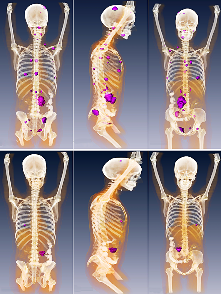
Authors: George Papadakis, MD, Ulas Bagci, PhD, Corina Millo, MD, Ingo Janssen, MD, Nicholas Patronas, MD, Constantine Stratakis, MD, D(med)Sci, and Karel Pacak, MD, PhD, DSc
Section on Medical Neuroendocrinology, PRAE, NICHD
Detection of metastatic paraganglioma with the novel imaging modality 68Ga-DOTATATE PET/CT compared with 18F-FDOPA PET/CT (frontal, lateral, and dorsal views)
Click image to enlarge.
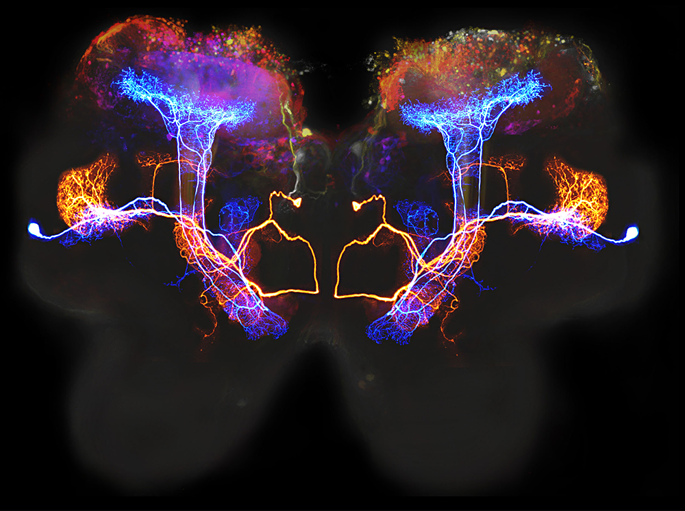
Authors: Nitin Gupta, PhD, and Mark Stopfer, PhD
Section on Sensory Coding and Neural Ensembles, PDN, NICHD
This image shows two pairs of β-lobe neurons (one colored here blue, one orange), which process olfactory information in the locust brain. Also visible in color (toward the top) are the mushroom bodies, brain areas associated with learning and memory. The β-lobe neurons were visualized by intracellular injection with neurobiotin. The mushroom body was visualized by mass fill with a dextran dye and imaged so that deeper parts are more blue and parts closer to the surface are red. The image is a composite of several different confocal pictures registered to the same locust brain (light gray).