

You are here: Home > Program in Perinatal Research and Obstetrics
Program in Perinatal Research and Obstetrics
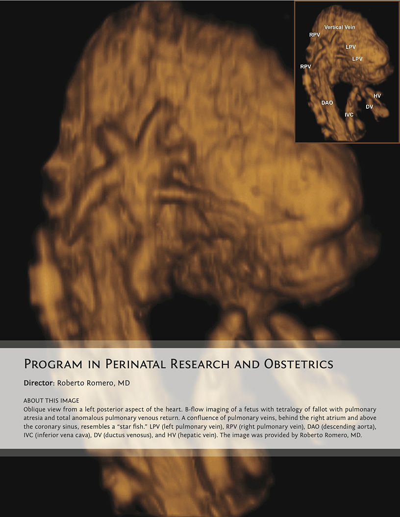
Director: Roberto Romero, MD
Click image to enlarge.
The Program in Perinatal Research and Obstetrics (PPRO) conducts clinical and laboratory research on maternal and fetal diseases responsible for excessive infant mortality in the United States. The Program focuses on the mechanisms of disease responsible for premature labor and delivery, with particular emphasis on the role of subclinical intrauterine infection and inflammation. The prenatal diagnosis of congenital anomalies is also a major area of interest.
The Perinatology Research Branch was created by Public Law (1993) to address the causes of the excessive infant mortality in the United States. The Branch, now called the Program in Perinatal Research in Obstetrics, uses a multidisciplinary approach to study complications of pregnancy, which include the disciplines of Maternal-Fetal Medicine, Neonatology, Placental Pathology, Perinatal Epidemiology, and High-Dimensional Biology (genomics, proteomics, metabolomics, and computational biology), as well as Immunology, Microbiology, and Nanomedicine. The Program is currently housed in Detroit, Michigan, on the campus of Wayne State University and at the Detroit Medical Center. The location is appropriate given the high perinatal and infant mortality in the city, as well as the contribution of ethnic social disparities to adverse pregnancy outcome. The PPRO provides state-of-the-art prenatal care to women enrolled in NICHD protocols and has made major contributions to the diagnosis of congenital anomalies and the understanding of mechanisms of disease in premature labor/delivery and preeclampsia.
Preterm birth is the leading cause of perinatal mortality and morbidity worldwide. The cost of prematurity in the U.S. alone is $26 billion per year. The Perinatology Research Branch proposed that preterm parturition is a syndrome caused by multiple pathologic processes, one of which is an untimely decline in progesterone action, which is manifested by silent shortening of the uterine cervix. Several lines of evidence suggest that administration of vaginal progesterone to patients with a short cervix may reduce the rate of preterm birth. Indeed, previous work conducted by the Program showed that asymptomatic women who have a cervical length of less than 15mm in the second trimester of pregnancy have a 50% likelihood of delivering an early preterm neonate. However, prediction of preterm birth should be accompanied by a strategy to reduce the frequency of this complication. The PPRO conducted a randomized clinical trial of vaginal progesterone vs. placebo in women with a short cervix and reported that treatment was associated with a 45% reduction in the rate of preterm birth (less than 33 weeks of gestation) and a reduction in the rate of respiratory distress syndrome, the most common complication of prematurity. The Branch continues to study mechanisms of disease responsible for spontaneous and indicated preterm birth (e.g., preeclampsia and intrauterine growth restriction) with the goal of identifying biomarkers to predict disease and therapeutic avenues for prevention, following the blueprint used for the prevention of preterm birth with universal cervical screening with ultrasound and vaginal progesterone administration to women with a short cervix.
Another major area of interest of the PPRO is the prenatal diagnosis of congenital anomalies. Congenital heart disease is the leading organ-specific birth defect as well as the leading cause of infant mortality from congenital malformations. More than half the infants afflicted with congenital heart disease are born to mothers without any previously known risk factors, which provides the impetus to perform a comprehensive screening examination of the fetal heart in all pregnancies. Imaging is a powerful instrument for scientific discovery and has changed the practice of obstetrics and maternal-fetal medicine. The single most important step that has made fetal medicine a discipline is the transformation of the fetus from an “invisible” to a visible subject through the use of ultrasound. This has allowed the definition of fetal anatomy, biometry, growth and the study of physiologic parameters, such as fetal cardiac function, sleep, and breathing.
Although ultrasound is the standard imaging modality in pregnancy, magnetic resonance imaging (MRI) has been used to characterize fetal anatomy when ultrasound cannot provide definitive diagnostic answers. MRI can provide unique information about fetal physiologic parameters (i.e., perfusion, oxygenation, and biochemistry) that are outside the domain of ultrasound. Moreover, MRI can be used to characterize the ontogeny of functional neuroconnectivity, as well as the potential relationship between insults that could alter neurodevelopment. Therefore, the goals of this project are to use quantitative MRI to gain insight into the anatomy, physiology, and pathology of the human fetus.