


You are here: Home > Program on Pediatric Imaging and Tissue Sciences
Program on Pediatric Imaging and Tissue Sciences
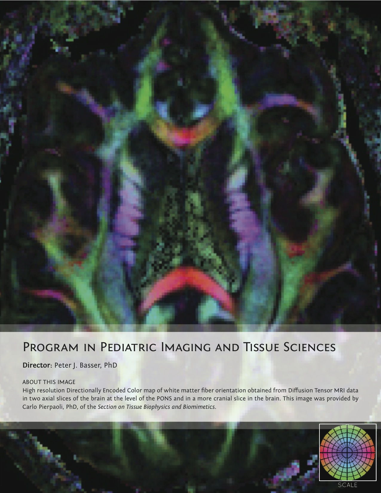
Director: Peter J. Basser, PhD
Click image to enlarge.
The new Program on Pediatric Imaging and Tissue Sciences (PPITS) was created in January 2010 to address critical, unmet needs in Pediatric Radiology and the basic sciences supporting it. This unique Program sponsors a broad range of basic, applied, and translationally oriented research activities aimed at improving the assessment of normal development and at the screening, diagnosis, or prognosis of diseases, disorders, or disabilities common in the pediatric population. To this end, PPITS scientists invent, develop, and apply non-invasive imaging methods and modalities employing quantitative imaging biomarkers that can sensitively and selectively identify key features of target tissues or organs. An essential and unique feature of this Program is that, to achieve these translational goals, PPITS supports and performs a broad range of basic and applied research in areas such as tissue sciences, aiming to identify and characterize potentially salient quantitative imaging biomarkers, as well as imaging physics and imaging sciences to permit their measurement.
Peter Basser heads the Section on Tissue Biophysics and Biomimetics, which strives to understand fundamental relationships between functional properties of soft tissues and their structure in vivo, in "engineered" tissue constructs, and in tissue analogs (e.g., polymer gels). This is achieved by studying complex biological processes and structure/function relationships in an integrative fashion, i.e., by probing a hierarchy of length and time scales, as well as developing an array of biological, mathematical, physical, and computational models and model systems. The Section produced a method based on anomalous X-ray scattering to measure the ion distribution around charged biopolymer molecules and built a tissue micro-osmometer that permits continuous monitoring of water uptake of small specimens. The Section also developed an experimental method to map the elastic properties of tissues and cells and designed and constructed calibration standards for diffusion MRI imaging. The Section designs and implements novel quantitative in vivo neuroimaging methods to scan children and adults noninvasively, combined with effective strategies for correcting distortion in diffusion-tensor imaging scans of the human brain. The Section elaborates new quantitative MRI methodologies to probe the microstructure and architectural organization of tissue, with an emphasis on the brain, including a method to measure the axon diameter distribution within nerve fascicles. Another MRI-based approach to characterize features of gray matter is intended to enable the architectonic (Brodmann) parcellation of the cerebral cortex in vivo.
The Section on Analytical and Functional Biophotonics, led by Amir Gandjbakhche, devises quantitative biophotonics methodologies and associated instrumentation to study biological phenomena at various length scales, from nanoscopic to microscopic. In the new Program, the Section is focusing on translation of technologies and methodologies to the bedside and the field. At the pre-clinical level, the Section is using a near-infrared, scanning, time-resolved imaging system to specifically quantify HER-2 receptors in tumor xenografts, with the goal developing a non invasive immune-imaging system. At the bedside, the Section 1) designed an optical camera, mounted in a conventional colposcope for active polarization imaging of cervical texture while improving statistical tools to enhance visualization of hidden structures (clinical protocol 09-CH-0180); 2) developed real-time algorithms to assess vascularity in AIDS-associated Kaposi's sarcoma (clinical protocol 08-CH-0001); 3) developed near-infrared spectroscopy (NIRS) as a noninvasive technique for measuring the local changes in cerebral hemodynamic levels associated with brain activity. The NIRS technique will be used on populations that include patients with traumatic brain injury (children, veterans), and autistic spectrum disorder patients. The ultimate goal of the project is to develop a small and portable near infrared instrument with novel neuro-imaging capabilities and potentially translate it to the bedside for use in hospital environments (clinical protocol 10-CH-0198).