

You are here: Home > Program on Pediatric Imaging and Tissue Sciences
Tissue Biophysics and Biomimetics

- Peter J. Basser, PhD, Head, Section on Tissue Biophysics and Biomimetics
- Ferenc Horkay, PhD, Staff Scientist
- Carlo Pierpaoli, MD, PhD, Staff Scientist
- Iren Horkayne-Szakaly, MD, Volunteer
- Candida Silva, PhD, Volunteer
- Michal Komlosh, PhD, Henry M. Jackson Foundation Contractor
- Evren Özarslan, PhD, Henry M. Jackson Foundation Contractor
- Amritha Nayak, Technical Intramural Research Training Awardee
- Lindsay Walker, MS, Guest Researcher
We attempt to understand fundamental relationships between function and structure in living tissues, "engineered" tissue constructs, and tissue analogs. Specifically, we are interested in how microstructure, hierarchical organization, composition, and material properties of tissue affect its biological function and dysfunction. We investigate biological and physical model systems at different time and length scales, making physical measurements in tandem with the development of mathematical and computational models. Primarily, we use water molecules to probe both equilibrium and dynamic interactions among tissue constituents over a wide range of time and length scales. To determine the equilibrium osmo-mechanical properties of well-defined model systems, we vary water content or ionic composition. To probe tissue structure and dynamics, we employ atomic force microscopy (AFM), small-angle X-ray scattering (SAXS), small-angle neutron scattering (SANS), static light scattering (SLS), dynamic light scattering (DLS), and nuclear magnetic resonance (NMR) relaxometry. We develop and use mathematical models to help us understand how observed changes in tissue microstructure and physical properties (e.g., mass, charge, and momentum) affect essential transport processes. The most direct noninvasive in vivo method for characterizing these transport processes in tissues is magnetic resonance imaging (MRI), which we use to follow microstructural changes in development, degeneration, aging, and trauma. A goal of our basic tissue sciences research is to translate our quantitative methodologies and the understanding we glean from them from "bench to bedside."
Virtual in vivo histology
This activity area has the dual goals of developing novel next-generation in vivo imaging methods to help us gain a better understanding of brain function and organization and to improve the diagnosis of neurological and developmental disorders. The most mature technology that we have so far invented and developed for these purposes is Diffusion Tensor MRI (DTI). With it, we measure a diffusion tensor of water, D, within an imaging volume, voxel-by-voxel. Information derived from D includes the local nerve fiber–tract orientation, the mean-squared distance that water molecules diffuse in each direction, the orientationally averaged mean diffusivity, and other intrinsic scalar (invariant) quantities (i.e., those that are independent of the laboratory coordinate frame used to make these measurements). The scalar parameters derived from D behave like quantitative histological "stains" obtained from endogenous tissue water, but are "developed" without exogenous contrast agents or dyes. The bulk or orientationally averaged diffusivity is the most successful imaging parameter proposed to date for identifying ischemic regions in the brain during acute stroke. Measures of diffusion anisotropy (e.g., the "fractional anisotropy" or FA) are widely used to follow changes in normally and abnormally developing white matter, including dysmyelination and demyelination. Our group also pioneered the use of diffusion tensor–derived color maps to encode nerve fiber orientation, maps that identify the main association, projection, and commissural white matter pathways in the brain. To assess anatomical connectivity among various functional brain regions, we also developed DTI "streamline" Tractography to track nerve fiber trajectories by continuously following the direction along which the apparent diffusivity is a maximal.
More recently, we invented and have been developing several advanced in vivo MR methods to measure fine-microstructural features of nerve fascicles, which previously could be measured by optical microscopy, and only by using laborious ex vivo histological methods. We are developing efficient in vivo methods for performing "k and q-space MRI" in the living brain. This approach can detect subtle microstructural and architectural features in both gray and white matter at a finer spatial resolution than DTI. We can also use this approach to characterize features of "anomalous" or fractal diffusion we recently observed in the neuropil. We recently developed a diffusion MRI method to measure the axon-diameter distribution within large white-matter fascicles, dubbing this method AxCaliber MRI. After careful validation studies, we recently reported the first in vivo measurement of axon diameter distributions within the corpus callosum in the rodent brain. This quantity is important neurophysiologically and developmentally, because axon diameter determines nerve conduction velocity and thus the rate of information transfer along nerve pathways. We are also developing mathematical models in tandem with these experiments to help discover important organizing principles to explain observed axon diameter distributions in different fascicles and features of information transfer between different brain regions.
While gray matter appears featureless in DTI, its microarchitecture is rich and varied, not only along the cortical surface, but within different cortical layers. We have been developing several noninvasive, in vivo methods to "drill down" into the image voxel to measure unique features of gray matter microstructure that are currently invisible in conventional MRI. One goal is to "parcellate" or segment the cerebral cortex in vivo into its distinct "Brodmann areas." To this end, we are developing advanced MRI sequences to probe correlations between microscopic displacements of water molecules in the neuropil as well as sophisticated mathematical models to infer distinguishing microstructural and morphological features of gray matter. We hope to use these new measurements to follow normal and abnormal development of the cerebral cortex noninvasively and in vivo.
Quantitative pediatric MRI
Traditionally, clinical MRI has relied on the acquisition of so called "weighted images" whose contrast is affected by many factors, some intrinsic to the tissue, and some dependent on the details of the experiment. The diagnostic utility of conventional MRI in many neurological disorders is unquestionable. However, the domain of conventional MRI is limited to revealing either gross morphological abnormalities or focal abnormalities resulting in regional differences in signal intensities within a given tissue. To detect pathology, conventional MRI relies on differences in contrast between areas that are supposedly affected and supposedly normal and is intrinsically insensitive to subtle global changes that may affect the entire tissue. Clinical MRI also lacks biological specificity. Although quantification per se does not assure such specificity, its use is necessary for developing imaging "biomarkers".
The MRI assessment of normal brain development and developmental disorders are greatly benefiting from "quantitative" clinical MRI techniques, in which one obtains maps of meaningful physical quantities or chemical variables that can be measured in physical units and compared among and between tissue regions, in longitudinal and multisubject studies. Quantitative MRI also increases sensitivity, providing a basis for monitoring subtle changes that occur during the progression or remission of disease by comparing measurements in a single subject with normative values acquired in a healthy population.
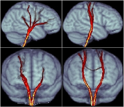
Click image to enlarge.
Figure 1. Diffusion Tensor MRI (DTI) based probabilistic tractography of the cortico-spinal tract
Diffusion imaging data can be distorted by several experimental artifacts, which can lead to false positives and false negatives in a connectivity analysis using Tractography methods. Left panels shows probabilistic tractography results from raw, uncorrected data. Right panels shows results after artifact correction. Tracts computed from uncorrected data reach anatomically incorrect brain regions such as frontal lobes but not the top cortical regions. Additionally, bilateral symmetry is absent. Stability, accuracy, and reproducibility in Tractography are greatly improved in the corrected data. Tractography is sensitive to artifacts that are generally disregarded. Care in data acquisition and processing is necessary to ensure robust connectivity analysis.
Our group is carrying out several clinical studies that utilize novel quantitative MRI acquisition and analysis methods aimed at improving accuracy and reproducibility in diagnosis, and detecting and following normal and abnormal development. These studies include:
- The NIH Study of Normal Brain Development, jointly sponsored by four NIH Institutes (NICHD, NIMH, NINDS, and NIDA) constitutes a multicenter study to advance our understanding of brain development in typical healthy children and in adolescents. Structural MR images and the results of standardized neuropsychological tests from this population are now available to researchers. Our role in this interdisciplinary project is to serve as a Diffusion Tensor MRI (DTI) Data-Processing Center (DPC). We have now processed all admissible DTI data, and the release of these data into a database accessible to all interested investigators is imminent. We are now analyzing the data, developing age-specific DTI atlases of normal brain development. We also released publicly the software we developed for this project and related documentation, which can be downloaded from http://www.tortoisedti.org. In particular, we are using our advanced DTI–processing pipeline to produce high-quality normative data from this project and to make them publicly available through the National Database for Autism Research (NDAR; http://ndar.nih.org).
- In collaboration with Susan Swedo, we are studying autistic subjects using DTI and quantitative relaxometry methods. Several MRI studies have reported abnormal features in the autistic brain, but no clear MRI "biomarker" of autism exists. The aim of this study is to use robust quantitative metrics to identify potential anatomical abnormalities in the autistic brain and identify candidate imaging biomarkers for this disorder.
- In collaboration with Kathy Warren, we are scanning children with pontine gliomas to identify MRI prognostic factors. With John Park, we are scanning children with supratentorial gliomas to differentiate recurrence from radiation necrosis.
- In collaboration with Filippo Arrigoni (of the Institute "Eugenio Medea," (http://www.emedea.it/english_medea), we are using multimodality MR imaging (DTI, fMRI, and quantitative relaxometry) to evaluate cerebral reorganization caused by different rehabilitation protocols in children with cerebral palsy and traumatic brain injury (TBI).
- In addition to these clinical studies, we are working on a Pilot grant under the auspices of the Center for Neuroscience and Regenerative Medicine (CNRM, http://www.usuhs.mil/cnrm) to develop enhanced software tools for analysis of diffusion MRI in traumatic brain injury (TBI) and posttraumatic stress disorder (PTSD). DTI provides essential information for the diagnosis of TBI and could be developed into an important tool for the assessment of potential structural damage in PTSD. The goal of this project is to provide a DTI data–processing pipeline to improve the accuracy and reproducibility of DTI findings for CNRM investigators and potentially the larger clinical and scientific community involved in TBI research. This will be accomplished by adding four new modules to our existing state-of-the-art DTI–processing pipeline as well as tools to enable calibration of DTI experiments using a new polymer-based diffusion MRI phantom we developed.
Looking forward, to permit analysis of novel MRI data sets, such as the one described above, as well as new clinical and biological applications of quantitative MRI, we need to create new mathematical, statistical, and image-sciences concepts and tools. To date, we have developed algorithms that generate a continuous, smooth approximation to the discrete, noisy, measured DTI field data to reduce noise and enable us to follow fiber tracts more reliably. We proposed a new Gaussian distribution for tensor-valued random variables that we used in designing optimal DTI experiments. In tandem, we developed nonparametric empirical (e.g., Bootstrap) methods for determining the statistical distribution of DTI–derived quantities in order to study, for example, the inherent variability and reliability of white matter fiber tract trajectories. These parametric and nonparametric statistical methods will enable us to apply powerful hypothesis tests to assess statistical significance in a wide range of important biological and clinical questions that are currently being addressed using ad hoc methods. We are also developing novel methods to generate group-average data or atlases from various subject populations. We are also developing methods for studying the reproducibility and reliability of tractography findings (Figure 1). We recently developed a diffusion MRI "phantom" to assure the quality of acquired MRI data in clinical and research sites worldwide. However, much work remains to be done to permit statistically significant inferences from clinical DTI data, particularly those obtained in longitudinal and multicenter studies.
Biopolymer physics: water-ion-biopolymer interactions

Click image to enlarge.
Figure 2. DNA condensation
Different configurations of DNA induced by a multivalent cationic polymer
The study of water-ion-polymer interactions has the potential to contribute to a more fundamental understanding of the ion-mediated structural organization of a plethora of charged biopolymers, particularly in physiological solution conditions. In polynucleic acids, proteins, and biological membranes, electrostatic interactions often play a dominant role in determining phase behavior. Charged macromolecules dissociate in solution forming macro-ions surrounded by an atmosphere of small mobile counterions. We study the structure and behavior of polyelectrolytes at different length and time scales using an array of scattering techniques in combination with macroscopic osmotic and mechanical methods. In addition, we perform modeling studies on polyelectrolyte solutions and hydrogels to determine the effect of ion concentration and ion valence on polymer morphology from the molecular scale to the nano-, micro-, and macroscales. Because of its ubiquity and importance in biological processes, we are particularly interested in understanding mechanisms of interactions between Ca2+ ions and charged biopolymers.
To help understand the nature of physical/chemical interactions in biomolecules and biomolecular assemblies, we have developed a comprehensive experimental approach to study their structure (morphology) and their thermodynamic properties in tandem as a function of the length scale (spatial resolution). This is achieved by combining macroscopic osmotic swelling-pressure and small-angle scattering measurements. Swelling-pressure measurements probe the system in the large length-scale range, providing information about its thermodynamic properties. High-resolution SANS and SAXS experiments allow us to investigate native biomolecules and relate changes in their physical properties (such as molecular conformation and osmotic pressure) with changes in environmental conditions (e.g., ion concentration, ion valance, pH, and temperature). Together, these measurements help us determine the length scales that govern or control the macroscopic thermodynamic properties of these molecules. This information cannot be obtained by other techniques.
We applied this approach to study the effect of multivalent cations, particularly Ca2+, on the structure of various biological model systems. Existing polyelectrolyte theories do not adequately describe the interactions of Ca2+ with charged macromolecules. Moreover, these interactions are difficult to study experimentally because above a low ion concentration threshold multivalent cations such as Ca2+ precipitate. By cross-linking charged biopolymers into gels, in which such macroscopic phase separation does not occur, we have extended the range of ion concentrations over which such systems remain stable and can be studied. In addition, gels mimic the crowded environment typically found in cells and tissue. In pilot studies, this new non-destructive procedure has been used to investigate cross-linked DNA and hyaluronic acid (HA) gels to determine the size of the structural elements that contribute to osmotic concentration fluctuations. We combined SANS and SAXS measurements to estimate the osmotic modulus of HA solutions in the presence of monovalent and divalent counterions. We determined the distribution of counterions around charged biopolymer molecules by using anomalous SAXS measurements. We identified different pathways to condense DNA molecules into nanoparticles (Figure 2), which have potential applications in therapeutic nanomedicine.
Functional properties of extracellular matrix (ECM)
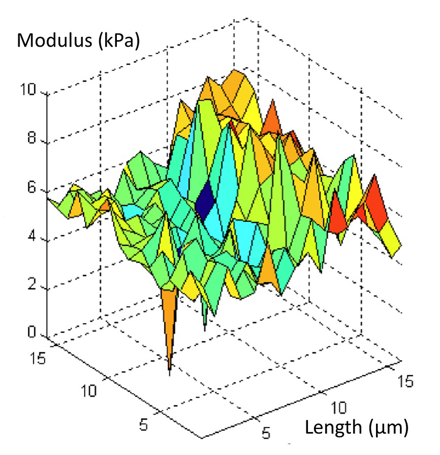
Click image to enlarge.
Figure 3. Elastic (Young's) modulus map of 1-day-old developing mouse cartilage
High-resolution map of local tissue stiffness obtained from AFM data and physical models
Our goal is to explain the physical basis of functional properties of cartilage and other ECMs in terms of their hierarchical structure, the matrices' components, and interactions among them. Understanding the physical and chemical mechanisms affecting cartilage swelling is essential to predicting load-bearing and lubrication, which are governed by osmotic and electrostatic forces that strongly depend on tissue hydration. Such understanding is also a prerequisite for the success of tissue engineering or regenerative medicine strategies, particularly for cartilage repair and regrowth.
One strategy we employ to gain a self-consistent physical picture of tissue structure/function relationships is to measure various physical/chemical properties of tissues and tissue analogs at different length and time scales by using a variety of complementary experimental techniques, (e.g., osmometry, SANS, SAXS, neutron spin-echo, SLS, DLS, AFM, and fluorescent light scattering).
The swelling behavior of cartilage is sensitive to both biochemical and microstructural changes occurring in development, disease, degeneration, and aging. To study the thermodynamics of cartilage hydration, an array of techniques is required that probe not only a wide range of length scales but are also a statistically representative volume of the tissue. Controlled hydration or swelling provides a direct means of determining functional properties of cartilage and of other ECMs. We have previously used controlled hydration of cartilage to measure physical/chemical properties of the collagen network and of the proteoglycans (PG) independently within cartilage.
We developed a novel tissue micro-osmometer to perform swelling experiments in a practical, rapid, and automated manner. This instrument can measure minute amounts of water absorbed by small tissue samples (less than 1 microgram) as a function of the equilibrium activity (pressure) of the surrounding water vapor. A quartz crystal detects the water uptake of a specimen attached to the crystal's surface. The high sensitivity of its resonance frequency to small changes in the amount of adsorbed water allows us to measure precisely the mass uptake of a tiny tissue specimen.
We used osmotic pressure measurements to determine the contributions of individual components of the ECM (e.g., aggrecan, HA, and collagen) to the total tissue swelling pressure. Our measurements on aggrecan/HA systems revealed that the osmotic modulus of the aggrecan–HA complex is enhanced compared with that of the random assemblies of aggrecan bottlebrushes, providing direct evidence that the aggrecan–HA complex raisses the load-bearing ability of cartilage. Our combined static and dynamic scattering measurements (SAXS, SANS, SLS, DLS, neutron spin-echo) demonstrated that aggrecan–HA assemblies exhibit microgel-like behavior and possess a remarkable insensitivity to changes in ionic environment, particularly to Ca2+ concentration. The results are consistent with aggrecan's role as an ion reservoir in cartilage and bone.
We developed a novel method for mapping the local elastic and osmotic properties of cartilage using AFM together with the tissue micro-osmometer. Many impediments that previously hindered the use of AFM to probe inhomogeneous samples, particularly biological tissues, were addressed by this new development. The technique utilizes the precise scanning capabilities of AFM to generate large volumes of compliance data from which the relevant elastic properties may be extracted (Figure 3). In conjunction with results obtained from high-resolution scattering measurements, micro-osmometry, and biochemical analysis, the technique allows us to map the spatial variations in the osmotic modulus within tissue specimens. Knowledge of the local osmotic properties of cartilage is particularly important, given that the osmotic modulus determines the compressive resistance to mechanical loading. We plan to apply these approaches in future studies of ECM regeneration and development.
Additional Funding
- Award G189AM from the Henry Jackson Foundation supports STBB's project in "AxCaliber MRI", which is under the joint auspices of the NIH, DoD, CNRM, and UHSUS.
- Award 60855 from the Henry Jackson Foundation supports STBB's project in "Radial Diffusion-weighted MRI Acquisitions for Diffusion Tensor imaging of TBI", which is under the joint auspices of the NIH, DoD, CNRM, and UHSUS.
- Award 305500-1.01-60855 from the Henry Jackson Foundation supports STBB's project in "Enhanced software tools for the analysis of diffusion MRI in TBI and PTSD", which is under the joint auspices of the NIH, DoD, CNRM, and UHSUS.
Publications
- Shemesh N, Özarslan E, Basser PJ, Cohen Y. Detecting diffusion-diffraction patterns in size distribution phantoms using double-pulsed field gradient NMR: theory and experiments. J Chem Phys 2010;132:034703-1-12.
- Horkay F, Basser PJ, Hecht AM, Geissler E. Counterion and pH-mediated structural changes in charged biopolymer gels. Macromol Symp 2010;291-292:354-361.
- Horkay F, Magda J, Alcoutlabi M, Atzet S, Zarembinski T. Structural, mechanical and osmotic properties of injectable hyaluronan-based composite hydrogels. Polymer 2010;51:4424-4430.
- Komlosh ME, Özarslan E, Lizak MJ, Horkay F, Schram V, Shemesh N, Cohen Y, Basser PJ. Pore diameter mapping using double pulsed-field gradient MRI and its validation using a novel glass capillary array phantom. J Magn Reson 2011;208:128-135.
- Walker L, Chang L-C, Koay CG, Sharma N, Cohen L, Verma R, Pierpaoli C. Effects of physiological noise in population analysis of diffusion tensor MRI data. NeuroImage 2011;54:1168-1177.
Collaborators
- Filippo Arrigoni, MD, Fondazione IRCCS Eugenio Medea, Bosisio Parini, Italy
- Yaniv Assaf, PhD, Tel Aviv University, Tel Aviv, Israel
- Alan Barnett, PhD, Clinical Brain Disorders Branch, NIMH, Bethesda, MD
- Preethi Chandran, PhD, Division of Bioengineering and Physical Science, NIBIB, Bethesda, MD
- Yoram Cohen, PhD, Tel Aviv University, Tel Aviv, Israel
- Emilios Dimitriadis, PhD, Division of Bioengineering and Physical Science, NIBIB, Bethesda, MD
- Raisa Freidlin, MS, Computational Bioscience and Engineering Laboratory, CIT, Bethesda, MD, NIH
- Erik Geissler, PhD, CNRS, Université Joseph Fourier de Grenoble, Grenoble, France
- Anne-Marie Hecht, PhD, CNRS, Université Joseph Fourier de Grenoble, Grenoble, France
- Stefano Marenco, PhD, Clinical Brain Disorders Branch, NIMH, Bethesda, MD
- Pedro Miranda, PhD, Universidade de Lisboa, Lisbon, Portugal
- Sinisa Pajevic, PhD, Mathematical and Statistical Computing Laboratory, CIT, NIH, Bethesda, MD
- John Park, MD, Surgical Neurology Branch, NINDS, Bethesda, MD
- Bradley Roth, PhD, Oakland University Department of Physics, MI
- Susan Swedo, MD, Pediatrics and Developmental Neuroscience Branch, NIMH, Bethesda, MD
- Kathy Warren, MD, Pediatric Oncology Branch, NCI, Bethesda, MD
- Brain Development Cooperative Group, Various
Contact
For more information, email pjbasser@helix.nih.gov or visit stbb.nichd.nih.gov.