

You are here: Home > Unit on Reproductive Endocrinology and Infertility
Reproductive Endocrinology and Infertility

- James Segars, MD, Head, Unit on Reproductive Endocrinology and Infertility
- Alicia Armstrong, MD, Associate Fellowship Program Director, Reproductive Endocrinology and Infertility
- William H. Catherino, MD, PhD, Special Volunteer
- Hisashi Koide, MD, PhD, Visiting Fellow
- Xiu-Ye Xing, MD, Visiting Postdoctoral Fellow
- Kimberly M. Baig, BS, MD/PhD Student
- Xiaoxiao C. Guo, Student Fellow
- Sydney Chang, BS, Clinical Research Training Program Fellow
- Soledad Jorge, BS, Clinical Research Training Program Fellow
- Ashley Lawler, BS, Howard Hughes Medical Institute Medical Student
The Unit on Reproductive Endocrinology and Infertility supports basic, translational, and clinical studies on reproductive disorders. Current research focuses on uterine leiomyomas (fibroids), endometriosis, health disparities, and reproductive disorders leading to infertility in women. We also support the accredited NICHD Clinical Fellowship Program in Reproductive Endocrinology and Infertility.
Uterine leiomyomas
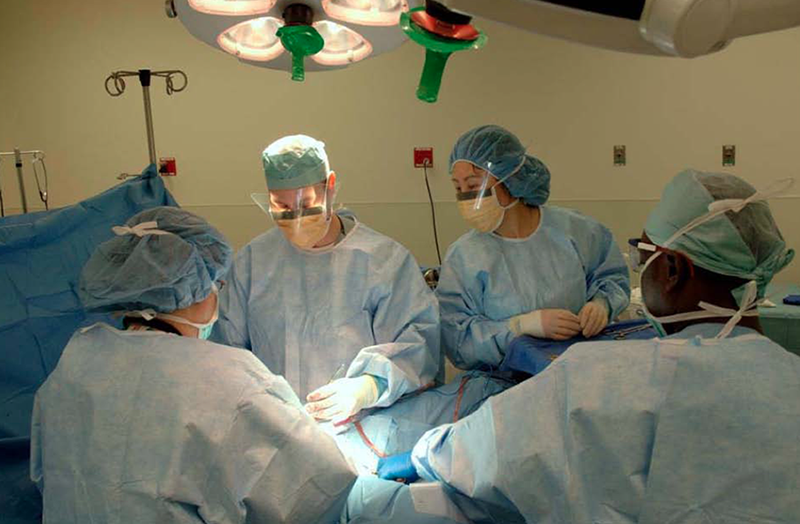
Click image to enlarge.
Figure 1. Surgeons in the Unit remove a symptomatic uterine leiomyoma.
The Unit on Reproductive Endocrinology conducts clinical, translational, and basic science studies of uterine leiomyoma.
Fifty percent of women in the United States develop uterine fibroids during their reproductive years, making the condition the most prevalent reproductive disorder of women. Despite its prevalence, the condition remains poorly understood. In the past year, we completed a cost estimate of the effect of uterine fibroids on the US health care system. We estimated that fibroids may represent a burden of up to $34 billion dollars annually, if adverse effects on reproduction are considered (1). One prominent feature of uterine fibroids is that the cells within the tumors produce a disordered and excessive extracellular matrix (ECM). In earlier work, we examined the characteristics of the cells that might lead to production of this excessive and fibrotic ECM and obtained evidence that mechanical signaling—a method of cell communication and activation—was attenuated in cells within a fibroid. During the past year, we examined surgical specimens and immortalized leiomyoma and myometrial cells and indeed found that mechanical signaling was lower in leiomyoma cells than in myometrial cells. Furthermore, given that anti-progestins cause reduction in fibroid size, we analyzed surgical specimens from patients treated with anti-progestins. Anti-progesterone–treated fibroids revealed alterations phosphorylation of proteins involved in mechanical signaling. Collectively, the findings suggest that altered mechanical signaling may contribute to leiomyoma growth (2).
Role of BRX (also known as AKAP13) in osmotic stress, inflammation, and development
Our previous studies on the gene BRX (now known as AKAP13), which we cloned, indicated that the gene encoded a large Rho-GEF proto-oncoprotein that is involved in estrogen and glucocorticoid receptor activation. In a recent collaborative study with Tomoshige Kino using mice haploinsufficient for BRX, we found that BRX was critical to the response of lymphocytes to osmotic stress through a pathway involving the nuclear factor of activated T cells 5 (NFAT5) and JIP4. In the past year, we reported that mice deficient for both alleles of the gene (Brx−/− mice) died in utero because of a failure of heart development. Our studies revealed that the BRX gene product coordinates Gas and Rho signaling with an essential transcription program in developing cardiomyocytes of mice, involving myocyte enhancer factor 2 C (MEF2C). In the coming year, we plan to examine the consequence(s) of haploinsufficiency for AKAP13 and to begin analysis of the Cre-Lox AKAP13 tissue–specific deletion in mice.
Assessment and preservation of ovarian function in women and girls undergoing cancer treatment
Because of effective chemotherapy for cancers, many young girls and women are now able to survive cancer, but find that reproductive function is impaired. Previously, we reported studies of a murine model of medical treatment prior to chemotherapy in an effort to explore the mechanisms responsible for preservation of ovarian function with gonadotropin releasing hormone (GnRH) antagonist therapy. We found that pre-treatment of mice with a GnRH antagonist reduced programmed cell death (apoptosis) in ovarian follicles associated with the chemotherapeutic agent cyclophosphamide. In the coming year, we plan to continue our clinical study of ovarian function in pediatric cancer survivors and have initiated additional studies on the mechanism involved in our murine model of GnRH−mediated chemo-protection.
Infertility and reproductive health disparities
We continued to conduct studies on infertility and reproductive health disparities. Previously, we examined health disparities in reproductive outcome in women undergoing assisted reproduction treatments. The clinical study indirectly examined the hypothesis that lower pregnancy outcomes observed in African-American women than in Caucasian women are a result of altered endometrial responses possibly due to higher levels of estrogen during the ovarian stimulation. Consistent with that hypothesis, when the endometrium was prepared similarly for embryo transfer, no ethnic differences in pregnancy rates were observed. In the past year, we examined factors such as cost in utilization of reproductive technologies by minority women. We found that economics and access to care influences use by African-American women but not by Hispanic women. Furthermore, despite increased use of assisted reproductive technologies by African American women, outcomes were worse than for Caucasian women. The results suggest that improved access to care may not translate into improved outcomes in some ethnic groups.
Publications
- Cardozo ER, Clark AD, Banks NK, Henne MB, Stegmann BJ, Segars JH. The estimated annual cost of uterine leiomyomata in the United States. Am J Obstet Gynecol 2012;206:211.e1-9.
- Norian JM, Owen CM, Taboas J, Korecki C, Tuan R, Malik M, Catherino WH, Segars JH. Characterization of tissue biomechanics and mechanical signaling in uterine leiomyoma. Matrix Biol 2012;31:57-65.
- Browne HN, Moon KS, Mumford SL, Schisterman EF, Decherney AH, Segars JH, Armstrong AY. Is anti-Müllerian hormone a marker of acute cyclophosphamide-induced ovarian follicular destruction in mice pretreated with cetrorelix? Fertil Steril 2011;96:180-186.
- McCarthy-Keith DM, Malik M, Britten J, Segars J, Catherino WH. Gonadotropin-releasing hormone agonist increases expression of osmotic response genes in leiomyoma cells. Fertil Steril 2011;95:2383-2387.
- Malik M, Segars J, Catherino WH. Integrin beta 1 regulates leiomyoma cytoskeletal integrity and growth. Matrix Biol 2012;E-pub ahead of print.
Collaborators
- Ayman Al-Hendy, MD, PhD, Meharry Medical College, Nashville, TN
- Paul Driggers, PhD, Uterine Fibroid Tissue Bank, USUHS-NICHD, Bethesda, MD
- Tomoshige Kino, PhD, Program in Reproductive and Adult Endocrinology, NICHD, Bethesda, MD
- Phyllis Leppert, MD, PhD, Duke University, Durham, NC
- Lawrence Nelson, MD, Program in Reproductive and Adult Endocrinology, NICHD, Bethesda, MD
- Lynnette Nieman, MD, Program in Reproductive and Adult Endocrinology, NICHD, Bethesda, MD
- Mark Payson, MD, Uniformed Services University of the Health Sciences, Bethesda, MD
- Pamela G. Robey, PhD, Craniofacial and Skeletal Diseases Branch, NIDCR, Bethesda, MD
- Rocky Tuan, PhD, University of Pittsburgh Medical School, Pittsburgh, PA
- Aradhana Venkatesan, MD, Radiology and Imaging Sciences, NIH Clinical Center, Bethesda, MD
- Heiner Westphal, MD, Program in Genomics of Differentiation, NICHD, Bethesda, MD
- Bradford J. Wood, MD, Center for Interventional Oncology, NIH Clinical Center, Bethesda, MD
Contact
For more information, email segarsj@mail.nih.gov.