

You are here: Home > Program on Pediatric Imaging and Tissue Sciences
Program on Pediatric Imaging and Tissue Sciences
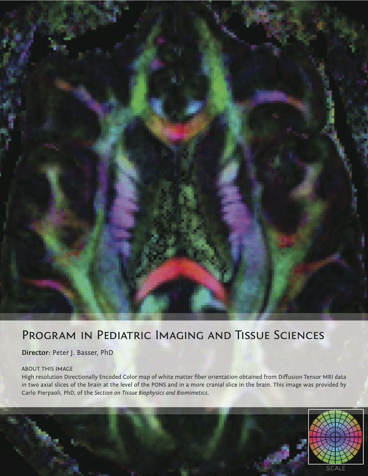
Director: Peter J. Basser, PhD
Click image to enlarge.
The Program on Pediatric Imaging and Tissue Sciences (PPITS) was created in January, 2010, to address critical, unmet needs in Pediatric Radiology and to provide a stronger scientific foundation supporting it. This unique Program sponsors a broad range of basic, applied, and translationally oriented research activities aimed at improving the assessment of normal development and at screening, diagnosis, and prognosis of diseases, disorders, or disabilities common in the pediatric population. To this end, PPITS scientists invent, develop, and apply non-invasive imaging methods and modalities to produce quantitative imaging biomarkers that can sensitively and selectively identify key features of target tissues or organs. An essential feature of this Program is that, to achieve these translational goals, PPITS supports and performs a wide array of basic and applied research in tissue sciences, which aim to identify and characterize potentially salient quantitative imaging biomarkers, as well as in the physical, mathematical, and imaging sciences to provide a conceptual framework for measuring such biomarkers.
Peter Basser heads the Section on Tissue Biophysics and Biomimetics, which strives to understand fundamental relationships between functional properties of soft tissues and their structure in vivo, in "engineered" tissue constructs, and in tissue analogs (e.g., polymer gels). Structure/function relationships are studied in an integrative fashion, primarily by probing key interactions and processes over a wide range of length and time scales, as well as by developing and studying relevant biological, mathematical, physical, and computational models and model systems. Recent achievements include a method based on anomalous X-ray scattering to measure the ion distribution around charged biopolymer molecules and construction of a tissue micro-osmometer that permits continuous monitoring of water uptake of small specimens. The Section also developed an experimental method to map the elastic properties of tissues and cells at a micron scale. STBB staff invent, develop, and translate novel quantitative in vivo methods for imaging tissues and organs, in particular, new quantitative MRI methodologies to probe tissue microstructure and architectural organization in the brain, and increasingly, in other soft tissues. Recent examples include non-invasive MRI methods to measure and map the diameter distribution of axons within nerve fascicles and to parcellate the cerebral cortex in vivo, based on microstructural features. From a clinical and translational perspective, STBB is involved in several MRI–based studies to migrate these promising new technologies from "bench to bedside."
The Section on Analytical and Functional Biophotonics, led by Amir Gandjbakhche, devises quantitative biophotonics methodologies and associated instrumentation to study biological phenomena at various length scales, from nanoscopic to microscopic. The Section focuses on the translation of technologies and methodologies to the bedside. At the pre-clinical level, the SAFB uses a near-infrared, scanning, time-resolved imaging system to quantify HER-2 receptors in tumor xenografts, with the goal developing a non-invasive immune-imaging system. At the "bedside" level, the Section (i) designed an optical camera, mounted in a conventional colposcope for active polarization imaging of cervical texture while improving statistical tools to enhance visualization of hidden structures (clinical protocol 09-CH-0180); (ii) developed real-time algorithms to assess vascularity in AIDS–associated Kaposi's sarcoma (clinical protocol 08-CH-0001); (iii) developed near-infrared spectroscopy (NIRS) as a noninvasive technique for measuring the local changes in cerebral hemodynamic levels associated with brain activity, a technique that will be used on patients with traumatic brain injury (children and veterans) or with autistic-spectrum disorder, with the goal of producing a small and portable near-infrared instrument with novel neuro-imaging capabilities, particularly for hospital use (clinical protocol 10-CH-0198).