

You are here: Home > Program on Pediatric Imaging and Tissue Sciences
Tissue Biophysics and Biomimetics

- Peter J. Basser, PhD, Head, Section on Tissue Biophysics and Biomimetics
- Ferenc Horkay, PhD, Staff Scientist
- Carlo Pierpaoli, MD, PhD, Staff Scientist
- Alan Barnett, PhD, Henry M. Jackson Foundation Contractor
- Okan Irfanoglu, PhD, Henry M. Jackson Foundation Contractor
- Michal Komlosh, PhD, Henry M. Jackson Foundation Contractor
- Cibu Thomas, PhD, Henry M. Jackson Foundation Contractor
- Alexandru Avram, PhD, Postdoctoral Fellow
- Elizabeth Hutchinson, PhD, Collaborating Scientist
- Ruiliang Bai, MS, Postbaccalaureate Visiting Fellow
- Dan Benjamini, MS, Postbaccalaureate Visiting Fellow
- Pooja Modi, BS, Postbaccalaureate Intramural Research Training Award Fellow
- Amritha Nayak, MS, Guest Researcher
We strive to understand fundamental relationships between function and structure in living tissues, “engineered” tissue constructs, and tissue analogs. Specifically, we are interested in how microstructure, hierarchical organization, composition, and material properties of tissues affect their biological function or dysfunction. We investigate biological and physical model systems at relevant hierarchical length and time scales, performing physical measurements in tandem with developing mathematical and computational models to explain salient features of these systems. Experimentally, we use water molecules to probe both equilibrium and dynamic interactions among tissue constituents from nanometers to centimeters and microseconds to lifetimes. To determine the equilibrium osmo-mechanical properties of well defined model systems, we vary water content or ionic composition systematically. To probe tissue structure and dynamics, we employ atomic force microscopy (AFM), small-angle X-ray scattering (SAXS), small-angle neutron scattering (SANS), static light scattering (SLS), dynamic light scattering (DLS), and one and two-dimensional nuclear magnetic resonance (NMR) relaxometry. We develop and use mathematical models to help us understand how observed changes in tissue microstructure and physical properties affect essential transport processes (e.g., of mass, charge, and momentum). The most direct and powerful noninvasive in vivo method for characterizing transport processes in tissues is magnetic resonance imaging (MRI), which we use to follow microstructural changes in development, degeneration, aging, and trauma. A goal of our basic tissue sciences research is to translate our bench-based quantitative methodologies, and the understanding we glean from them, to the bedside.
Virtual in vivo histology
Click image to enlarge.
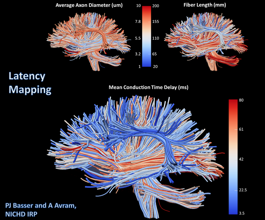
Figure 1. First in vivo human "latency map"
MRI Latency Imaging or Mapping is a non-invasive method for inferring an important feature of "functional connectivity" in the brain from in vivo MRI measurements of "structural connectivity." Specifically, we use diffusion MRI "Tractography" to obtain the lengths of white matter pathways connecting different gray matter Brodmann-like "areas" in the brain (top right); another MRI measurement technique we also invented measures mean axon diameters along those pathways (top left). From them, we can estimate the conduction delays or "latencies" between different brain gray matter areas using well-known neurophysiological scaling laws for myelinated axons, which relate axon diameter and conduction velocity (bottom center). The image shows our first in vivo MRI latency maps, obtained in a normal volunteer, in which latencies along different white matter pathways are encoded by color. In vitro experiments are planned to validate latency estimates obtained by this method.
We aim to develop novel next-generation in vivo MRI methods to better understand brain function and organization and to improve the diagnosis of neurodevelopmental disorders. To these ends, the most mature technology that we invented and developed is Diffusion Tensor MRI (DTI). With it, we measure a diffusion tensor of water, D, voxel-by-voxel within an imaging volume. Information derived from D includes the local nerve fiber–tract orientation, the mean-squared distance that water molecules diffuse in each direction, the orientationally averaged mean diffusivity, and other intrinsic scalar (invariant) quantities (i.e., those that are independent of the laboratory coordinate frame used to make these measurements). The scalar parameters derived from D behave like quantitative histological “stains" even though they are obtained from endogenous tissue water and are “developed” without exogenous contrast agents or dyes. The bulk or orientationally averaged diffusivity is the most successful DTI parameter proposed to date for identifying ischemic regions in the brain during acute stroke. Our measures of diffusion anisotropy (e.g., the "fractional anisotropy" or FA) are widely used to follow changes in normally and abnormally developing white matter, including dysmyelination and demyelination. Our group also pioneered the use of fiber direction–encoded color (DEC) maps (derived from D) to display the orientation of the main association, projection, and commissural white matter pathways in the brain. To assess anatomical connectivity among various functional brain regions, we also developed DTI "Streamline" Tractography to track white matter trajectories by continuously following the direction along which the apparent diffusivity is maximal.
More recently, we invented and have been developing more advanced in vivo MR methods to measure fine-microstructural features of axons and fascicles, which previously could be measured only optically using laborious ex vivo microhistological methods. We are developing efficient means for performing "k and q-space MRI" in the living brain, the most recent of which is "Mean Apparent Propagator" (MAP) MRI. The approach can detect subtle microstructural and architectural features in both gray and white matter at a finer spatial resolution than can DTI. We can also use MAP-MRI to characterize features of "anomalous" or fractal diffusion that we recently observed in neuropil (consisting of a patchwork of interwoven dendrites and axons and of neuroglial cells in the gray matter of the central nervous system). We also developed a diffusion–MRI method to measure the axon-diameter distribution (ADD) within large white-matter fascicles, dubbing it AxCaliber MRI. After careful validation studies, we reported the first in vivo measurement of ADDs within the rodent corpus callosum. The ADD is important neurophysiologically and developmentally given that axon diameter determines nerve conduction velocity and therefore affects the rate of information transfer along nerve pathways and among various brain regions. We developed mathematical models in tandem with these experiments to help explain observed ADDs in different fascicles that maximize information flow along them while minimizing metabolic demands. We also developed novel multiple pulsed-field gradient (mPFG) methods and demonstrated their feasibility for use in vivo on conventional clinical MRI scanners. The methods provide an independent measurement of mean axon diameter and other features of cell size and shape.
While gray matter appears featureless in DTI maps, its microarchitecture is rich and varied, not only along the brain's cortical surface but also within and among its different cortical layers. We have been developing several noninvasive, in vivo methods to "drill down" into the image voxel to measure unique features of cortical gray matter microstructure that are currently invisible in conventional MRI. One goal is to "parcellate" or segment the cerebral cortex in vivo into its distinct cytoarchitechtonic or Brodmann-like areas. To this end, we are developing advanced MRI sequences to probe correlations among microscopic displacements of water molecules in the neuropil as well as sophisticated mathematical models to infer distinguishing microstructural and morphological features of gray matter. We continue to develop such methods, which will permit us to follow normal and abnormal development of the cerebral cortex noninvasively and in vivo.
Quantitative pediatric MRI
MRI is considered safer for noninvasively scanning pediatric subjects than X-ray–based methods, such as computed tomography (CT). However, clinical MRI still lacks the quantitative character of CT data. Clinical MRI relies upon the acquisition of "weighted images," whose contrast is affected by many factors, some intrinsic to the tissue, and some dependent on the details of the experiment and experimental design. The diagnostic utility of conventional MRI in many neurological disorders is unquestionable. However, the scope of conventional MRI applications is limited to revealing either gross morphological or focal abnormalities, which result in regional differences in signal intensities within a given tissue. To detect pathology, conventional MRI relies on differences in contrast between areas that are presumed "affected" and presumed "normal", rendering it intrinsically insensitive to subtle global changes that may affect the entire tissue or organ. Clinical MRI also lacks biological specificity. Although quantification per se does not assure specificity, it is nonetheless necessary for developing imaging "biomarkers." In particular, the MRI assessment of normal brain development and developmental disorders has benefiting greatly from "quantitative" clinical MRI techniques, in which one obtains maps of meaningful physical quantities or chemical variables that can be measured in physical units and compared among and between tissue regions, in both longitudinal and cross-sectional studies. Here, quantitative MRI also increases sensitivity, providing a basis for monitoring subtle changes that occur during the progression or remission of disease by comparing measurements in a single subject against normative values acquired in a healthy population. Quantitative MRI methods should also enhance our ability to perform comparative effectiveness research for a variety of diagnosis and therapy assessments.
Our group is carrying out several clinical studies that utilize novel quantitative MRI acquisition and analysis methods, whose aim is to improve accuracy and reproducibility in diagnosis and to detect and follow normal and abnormal development. The studies include the following:
- The NIH Study of Normal Brain Development, jointly sponsored by four NIH Institutes (NICHD, NIMH, NINDS, and NIDA), is a multi-center effort to advance our understanding of normal brain development in typical healthy children and in adolescents. The Brain Development Cooperative Group (http://www.brain-child.org), created by this mechanism, is still active, publishing numerous papers each year. Structural MRIs and the results of standardized neuropsychological tests performed on this population are available to researchers. Our role in this interdisciplinary project has been to serve as the DTI Data-Processing Center (DPC). While we have now processed all admissible DTI data and released them to a database accessible to all interested investigators, we continue to analyze the data, developing age-specific DTI atlases of normal brain development. We also released the software we developed for this project publicly (and related documentation), which can be downloaded from http://www.tortoisedti.org. We continue to support and update the software. We are continuing to use our advanced DTI–processing pipeline to produce high-quality normative data from this project and to make them publicly available through the National Database for Autism Research (NDAR; nda.nih.gov).
- In collaboration with Susan Swedo, we study autistic subjects using DTI and quantitative relaxometry methods. Several MRI studies reported abnormal features in the autistic brain, but no clear MRI "biomarker" of autism exists. The aim of this study is to use robust quantitative metrics to identify potential anatomical abnormalities in the autistic brain and to find candidate imaging biomarkers for the disorder (an earlier study was described in Walker et al., Biol Psychiatry 2012;72:1043).
- In collaboration with Katherine Warren, we acquire quantitative MRI data in children with pontine gliomas to identify MRI prognostic factors. With John Park, we are scanning subjects with supratentorial gliomas to distinguish between recurrence and radiation necrosis.
- In collaboration with Filippo Arrigoni (http://www.emedea.it/english_medea), we use multimodal MR imaging (DTI, fMRI, and quantitative relaxometry) to evaluate cerebral reorganization caused by various rehabilitation protocols in children with cerebral palsy and traumatic brain injury (TBI). We collected diffusion MRI data on subjects affected by hereditary spastic paraparesis with the pure form of the disease, as well as those with additional cognitive impairment. There are remarkable neuroanatomic differences between the two groups.
- We are working under the auspices of the Center for Neuroscience and Regenerative Medicine (CNRM, http://www.usuhs.mil/cnrm) to investigate potential plasticity changes after rehabilitation in military personnel affected by TBI and post-traumatic stress disorder (PTSD).
- In collaboration with Sharon Juliano, we received support from the Congressionally Directed Medical Research Program (CDMRP) to investigate TBI in the ferret, using advanced MRI developed within STBB and histopathology techniques provided by Dr. Juliano's laboratory. The proposal is funded, and we are currently acquiring MRI data.
Our involvement in TBI research is expanding. DTI provides essential information for the diagnosis of TBI and could be developed into an important tool for the assessment of potential structural damage in PTSD. For clinical applications, however, reliable imaging protocols are needed. Part of our work is to develop a robust DTI data–processing pipeline in order to improve the accuracy and reproducibility of DTI findings for Center for Neuroregenerative Medicine (CNRM) investigators and for the larger clinical and scientific community involved in TBI research. To this end, we are adding new modules to our existing state-of-the-art DTI data–processing pipeline as well as tools to permit calibration of DTI experiments, using our novel polymer-based diffusion MRI phantom that we developed and are disseminating to clinical sites.
Looking ahead, to permit analysis of novel MRI data sets such as those described above, as well as to develop new clinical and biological applications of quantitative MRI, we need to create a new mathematical, statistical, and image sciences–based infrastructure. To date, we have developed algorithms that generate a continuous, smooth approximation of the discrete, noisy, measured DTI field data so as to reduce noise and allow us to follow fiber tracts more reliably. We proposed a novel Gaussian distribution for tensor-valued random variables that we used in designing optimal DTI experiments and interpreting their results. In tandem, we developed nonparametric empirical (e.g., Bootstrap) methods for determining the statistical distribution of DTI–derived quantities in order to study, for example, the inherent variability and reliability of white-matter fiber-tract trajectories. Such parametric and nonparametric statistical methods will enable us to apply powerful hypothesis tests to assess statistical significance in a wide range of important biological and clinical questions that are currently being addressed using ad hoc statistical methods. We are also developing novel methods to generate group-average data or atlases from various subject populations. Recently, our group has been developing methods for studying the reproducibility and reliability of different tractography methods, given that, increasingly, the methods are being used to assess anatomical connections between different brain regions in vivo. However, much work remains to be done in order to remedy MRI artifacts and permit one to draw statistically significant inferences from clinical DTI data, particularly from those obtained in longitudinal and multi-center studies.
Biopolymer physics: water-ion-biopolymer interactions
Molecular self-organization is ubiquitous in biology. Examples include DNA nanostructure formation and protein folding. Self-assembly of aberrant proteins into nanofibers is associated with neurodegenerative conditions, such as Alzheimer’s, Pick’s, Parkinson’s, and Huntington’s disease. One objective of this research is to better understand self-assembly from a physical perspective by studying, primarily in polynucleic acids and proteins, particularly the interactions between water, ions, and the biopolymer.
By combining macroscopic osmotic swelling pressure measurements and small-angle scattering measurements, we developed a multi-scale experimental approach to study the structure (morphology) and thermodynamic interactions in biopolymer assemblies as a function of the length scale (spatial resolution). Swelling pressure measurements probe the system at large (macroscopic) length scales, providing information on its overall thermodynamic response, while SANS and SAXS allow us to investigate biopolymers at the microscopic level. Measurements of physical properties, such as molecular conformation and osmotic pressure, as a function of changes in environmental conditions, such as ion concentration, ion valance, pH, and temperature, help us uncover which length scales govern key macroscopic thermodynamic properties. The information cannot be obtained by other techniques.
Divalent cations, particularly calcium ions, are ubiquitous in the biological milieu. However, existing theories of polyelectolyte solutions do not adequately explain or account for their effects on, or interactions with, charged macromolecules. In pilot studies, we used our new non-destructive procedure to determine the effect of calcium ions on the physical properties of hyaluronic acid (HA) and DNA gels. The gel systems mimic certain essential features of the extracellular matrix (ECM) and have potential as scaffolds for tissue-regeneration applications. We investigated the binding mechanism in glucose sensors made from smart zwitterionic hydrogels containing boronic acid moieties. Our combined SANS and osmotic pressure measurements provided a thermodynamic explanation for the enhanced selectivity of the gels for glucose over fructose. This class of material is important for the development of implantable continuous glucose sensors for use in Type I and Type II diabetes.
We also developed a method to control the size, compactness, and stability of DNA nano-particles by mediating their interactions with ions. We quantified the effects of salt, pH, and temperature on the stability of the nanoparticles and on their biological activity. Such polyplexes are pathogen-like particles, 70–300 nm in size, and with shapes resembling spherical viruses that evolved naturally to deliver nucleic acids to cells. The nano-particles contain the pDNA in an interior surrounded by a synthetic polymer bearing surface sugar residues, which are recognized by "M" cells and dendritic cells as pathogens. The biological activity of such nano-medicines depends on the competing requirements of sufficient stability to escape endosomal degradation after cellular uptake with retention of biological activity upon arrival in the nucleus, where the nano-particle releases the pDNA to achieve gene expression. Such knowledge is essential to design and control properties of DNA–based nano-medicines. DNA–based vaccines are promising in several disease indications and have potential applications in the treatment of infectious diseases, cancer, and allergies.
We studied nanostructures formed from small gelator molecules via enzyme-assisted self-assembly inside live cells. The gelator molecule consists of a self-assembly motif and an enzyme (phosphatase) substrate (tyrosine phosphorous ester). After the enzymatic reaction removes the phosphorous group, intermolecular interactions facilitate the self-assembly of the molecules, and they form nanofibers. The approach offers a new way to control cell behavior. For example, intracellular formation of molecular assemblies could be used to selectively inhibit the growth of cells that over-express certain enzymes. Developing molecular self-assembly–based approaches and exploring their potential applications in biomedicine (e.g., intracellular drug delivery) may ultimately lead to new ways to regulate cell functions.
Functional properties of extracellular matrix
Our goal is to understand and quantify the interactions among the major macromolecular components of extracellular matrix (ECM) that give rise to their key functional properties. Specifically, we study thermodynamic interactions among collagen, proteoglycans, water, and ions that govern their biomechanical and transport properties, using cartilage as a model system.
The biomechanical behavior of cartilage and other ECMs reflects both biochemical and microstructural changes occurring during development, disease, degeneration, and aging. Understanding the basis of key functional properties of cartilage requires an array of experimental techniques that probe a wide range of relevant length and time scales. Knowledge of the physical and chemical mechanisms affecting cartilage swelling (hydration) is essential for predicting its load-bearing and lubricating abilities, which are mainly governed by osmotic and electrostatic forces. The knowledge can aid in tissue-engineering or regenerative-medicine strategies to grow, repair, and reintegrate replacement cartilage. To gain a self-consistent physical picture of tissue structure/function relationships, we measure various physical/chemical properties of tissues and tissue analogs at different length- and time-scales using a variety of complementary static and dynamic experimental techniques, e.g., osmometry, SANS, SAXS, neutron spin-echo (NSE), SLS, DLS, AFM, and fluorescence correlation spectroscopy (FCS).
Controlled tissue hydration provides a direct means of determining the patency and load-bearing ability of cartilage. Previously, we used controlled hydration to measure physical/chemical properties of the collagen network and of proteoglycans (PG). We developed and built a novel tissue micro-osmometer to perform swelling pressure measurements in a practical, rapid, and eventually automated manner. The instrument measures minute amounts of water vapor absorbed by small tissue samples (less than 1 microgram) as a function of the water activity (vapor pressure). A quartz crystal detects the water uptake of the tissue specimen. The high sensitivity of the quartz crystal's resonance frequency to small changes in the amount of adsorbed water allows us to measure mass uptake precisely.
We use osmotic pressure measurements to determine the contributions of individual components of cartilage ECM (e.g., aggrecan, hyaluronic acid [HA], and collagen) to the total tissue-swelling pressure. Our measurements on aggrecan–HA systems revealed that, in the physiological concentration range, the osmotic modulus of the aggrecan–HA complex exceeds that of random assemblies of aggrecan bottlebrushes, providing direct evidence that the aggrecan–HA complex increases the load-bearing ability of cartilage. In addition, we demonstrated that aggrecan–HA assemblies behave like microgels, contributing to improved tissue-lubricating ability. We also found that aggrecan is highly insensitive to changes in the ionic environment, particularly to Ca2+ ions. The latter result is consistent aggrecan's role as a calcium ion reservoir mediating calcium metabolism in cartilage and bone.
We also developed a novel method for mapping the local elastic properties of cartilage (and other heterogeneous tissues) using AFM. Many impediments that previously hindered AFM's use to probe biological samples were addressed. The technique utilizes the precise scanning capabilities of AFM to generate compliance "maps," from which the relevant elastic properties can be extracted. We combined AFM with tissue micro-osmometry to produce elastic and osmotic modulus maps of cartilage. The results clearly show that the spatial variation of the osmotic modulus is similar to that of the elastic (shear) modulus but that the modulus's numerical value is significantly greater. Knowledge of the osmotic modulus is particularly important because it determines the tissue's resistance to external compressive loads.
Additional Funding
- Award 305500-1.01-60855 from the Henry Jackson Foundation supports STBB's project in "Enhanced Software Tools for the Analysis of Diffusion MRI in TBI and PTSD," which is under the joint auspices of the NIH, DoD, CNRM, and USUHS.
- Award 306135-2.01-60855 from the Henry Jackson Foundation supports STBB's project "Clinical Double Pulsed-Field Gradient (dPFG) MRI for Mild TBI Assessment," which is under the joint auspices of the NIH, DoD, CNRM, and USUHS.
Publications
- Komlosh ME, Özarslan E, Lizak MJ, Horkayne-Szakaly I, Freidlin RZ, Horkay F, Basser PJ. Mapping average axon diameters in porcine spinal cord white matter and rat corpus callosum using d-PFG MRI. Neuroimage 2013;78:210–216.
- Özarslan E, Koay CG, Shepherd TM, Komlosh ME, Irfanoglu MO, Pierpaoli C, Basser PJ. Mean Apparent Propagator (MAP) MRI: a novel diffusion imaging method for mapping tissue microstructure. Neuroimage 2013;78:16-32.
- Gao Y, Kuang Y, Du X, Zhou J, Chandran P, Horkay F, Xu B. Imaging self-assembly dependent spatial distribution of small molecules in a cellular environment. Langmuir 2014;29:15191−15200.
- Eliav U, Komlosh ME, Basser PJ, Navon G. Collagen composition and content-dependent contrast in porcine annulus fibrosus achieved by using double quantum and magnetization transfer filtered UTE MRI. Magn Reson Med 2014;71(1):388-393.
- Irfanoglu MO, Modi P, Nayak A, Knutsen A, Sarlls J, Pierpaoli C. DR-BUDDI: diffeomorphic registration for blip up-down diffusion imaging. Med Image Comput Comput Assist Interv 2014;17(1):218-226.
Collaborators
- Filippo Arrigoni, MD, Fondazione IRCCS Eugenio Medea, Bosisio Parini, Italy
- Alan Barnett, PhD, Clinical Brain Disorders Branch, NIMH, Bethesda, MD
- Madison Berl, PhD, Children's National Medical Center, Washington, DC
- Preethi Chandran, PhD, Howard University, Washington, DC
- Yoram Cohen, PhD, Tel Aviv University, Tel Aviv, Israel
- Emilios Dimitriadis, PhD, Division of Bioengineering and Physical Science, NIBIB, Bethesda, MD
- Uzi Eliav, PhD, Tel Aviv University, Tel Aviv, Israel
- Raisa Freidlin, PhD, Computational Bioscience and Engineering Laboratory, CIT, NIH, Bethesda, MD
- Erik Geissler, PhD, CNRS, Université Joseph Fourier de Grenoble, Grenoble, France
- Anne-Marie Hecht, PhD, CNRS, Université Joseph Fourier de Grenoble, Grenoble, France
- Iren Horkayne-Szakaly, MD, Armed Forces Institute of Pathology, Washington, DC
- Sharon Juliano, PhD, Uniformed Services University of the Health Sciences, Bethesda, MD
- Stefano Marenco, PhD, Clinical Brain Disorders Branch, NIMH, Bethesda, MD
- Pedro Miranda, PhD, Universidade de Lisboa, Lisbon, Portugal
- Gil Navon, PhD, Tel Aviv University, Tel Aviv, Israel
- Uri Nevo, PhD, Tel Aviv University, Tel Aviv, Israel
- Evren Özarslan, PhD, Brigham and Womens Hospital, Boston, MA
- Sinisa Pajevic, PhD, Mathematical and Statistical Computing Laboratory, CIT, NIH, Bethesda, MD
- John Park, MD, PhD, Surgical Neurology Branch, NINDS, Bethesda, MD
- Bradley Roth, PhD, Oakland University, Rochester, MI
- Susan Swedo, MD, Pediatrics and Developmental Neuroscience Branch, NIMH, Bethesda, MD
- Katherine Warren, MD, Pediatric Oncology Branch, Center for Cancer Research, NCI, Bethesda, MD
- Brain Development Cooperative Group, Various
Contact
For more information, email pjbasser@helix.nih.gov or visit stbb.nichd.nih.gov or neuroscience.nih.gov/Faculty/Profile/peter-basser.aspx.