

You are here: Home > Section on Cellular and Synaptic Physiology
Hippocampal Interneurons and Their Role in the Control of Network Excitability

- Chris J. McBain, PhD, Head, Section on Cellular and Synaptic Physiology
- Kenneth Pelkey, PhD, Staff Scientist
- Ramesh Chittajulla, PhD, Senior Research Fellow
- Gulcan Akgul, PhD, Postdoctoral Fellow
- Elizabeth Barksdale, PhD, Postdoctoral Fellow
- Michael Craig, PhD, Postdoctoral Fellow
- Jason Wester, PhD, Postdoctoral Fellow
- Megan Wyeth, PhD, Postdoctoral Fellow
- Xiaoqing Yuan, MSc, Biologist
- Geoff Vargish, BS, Graduate Student
- Steven Hunt, Biologist
Click image to enlarge.
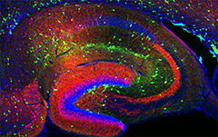
Figure 1. Interneurons in the GAD-65 GFP mouse
Numerous subpopulations are present within a hippocampal section derived from a GAD-65 GFP–transgenic mouse (GAD-65, the enzyme that catalyzes the synthesis of GABA, is a glutamic acid decarboxylase).
Cortical and hippocampal local-circuit GABAergic inhibitory interneurons are “tailor-made” to control Na+- and Ca2+-dependent action potential generation, regulate synaptic transmission and plasticity, and pace large-scale synchronous oscillatory activity. The axons of this diverse cell population make local, usually short-range projections (some subpopulations project their axons over considerable distances) and release the inhibitory neurotransmitter gamma-aminobutyric acid (GABA) onto a variety of targets. A mounting appreciation of the roles played by interneurons in several mental health conditions such as epilepsy, stroke, Alzheimer's disease, and schizophrenia have placed this important cell type center stage in cortical circuit research. Our main objective is to understand the developmental programs that regulate their integration into cortical circuits and how both ionic and synaptic mechanisms regulate the activity of cortical neurons at the level of small, well-defined networks. To this end, we use a variety of electrophysiological, immunohistochemical, molecular, and genetic approaches in both wild-type and transgenic animals. Over the past five years, we have continued our study of the differential mechanisms of glutamatergic and GABAergic synaptic transmission and plasticity within the hippocampal formation and the modulation of voltage- and ligand-gated channels expressed in inhibitory neurons. We also incorporated genetic approaches to unravel the embryogenesis and development of hippocampal interneurons and the circuits in which they are embedded. We are particularly interested in discovering the rules that dictate co-ordinated protein expression in nascent interneuron subpopulations as they migrate and integrate into the developing cortical circuit.
Dual embryonic origins of functionally distinct hippocampal O-LM cells revealed by differential expression of the serotonin receptor 5-HT3AR
For network synchronization and rhythmogenesis, forebrain circuits rely on a relatively small but remarkably diverse population of GABAergic interneurons to bind to and entrain large principal-cell assemblies. Despite the high degree of heterogeneity across cortical interneurons, members of a given subtype typically exhibit homogenous developmental origins, neuromodulatory response profiles, morphological characteristics, neurochemical signatures, and electrical features. We found a surprising divergence amongst hippocampal oriens-lacunosum moleculare (O-LM) projecting interneurons that were hitherto considered a homogeneous cell population. Combined immunocytochemical, anatomical, and electrophysiological interrogation of transgenic mice with GFP-labeled serotonin receptors revealed that O-LM cells parse into caudal ganglionic eminence–derived 5-HT3AR–expressing, and medial ganglionic eminence–derived 5-HT3AR–lacking subpopulations. The two cohorts differentially participate in network oscillations, with 5-HT3AR–containing O-LM cell recruitment dictated by serotonergic tone. Thus, members of a seemingly uniform interneuron population can exhibit distinct circuit functions and neuromodulatory properties dictated by disparate developmental origins.
Developmental origin dictates interneuron AMPA and NMDA receptor subunit composition and plasticity.
Disrupted maturation of excitatory synapse in cortical GABAergic interneurons can precipitate an imbalance in circuit excitation/inhibition that is thought to underlie the progression of neuropsychiatric disorders such as schizophrenia. However, establishing the basic developmental programs for nascent synapses in GABAergic cells is confounded by their sparsity, heterogeneity, and the late postnatal acquisition of subtype-defining characteristics. To overcome these hurdles, we investigated the synaptic properties of individual interneurons throughout early postnatal development, targeting cells based on lineage from medial or caudal ganglionic eminence (MGE and CGE) progenitors and thus enabling interrogation of non-overlapping interneuron populations whose fates can be predicted from origin. We found stereotyped developmental differences in the synaptic glutamate receptors AMPAR and NMDAR between MGE– and CGE–derived interneurons, including differential GluN2 subunit composition of NMDARs and expression of Ca2+-permeable AMPARs. Our findings establish ganglionic eminence–dependent rules for early postnatal synaptic integration programs of distinct interneuron cohorts including parvalbumin- and cholecystokinin-expressing basket cells.
5-HT1A receptor activation hyperpolarizes pyramidal cells and suppresses hippocampal gamma oscillations via Kir3–channel activation: relevance for schizophrenia.
Rhythmic cortical neuronal oscillations in the gamma frequency band (30–80 Hz, gamma oscillations) have been associated with cognitive processes such as sensory perception and integration, attention, learning, and memory. Gamma oscillations are disrupted in disorders for which cognitive deficits are hallmark symptoms, such as schizophrenia and Alzheimer's disease. In vitro, various neurotransmitters have been found to modulate gamma oscillations. Serotonin (5‐HT) has long been known to be important for both behavioral and cognitive functions such as learning and memory. Multiple 5‐HT receptor subtypes are expressed in the CA3 region of the hippocampus, and high doses of 5‐HT reduce the power of induced gamma oscillations. Hypothesizing that 5‐HT may have cell‐ and receptor subtype–specific modulatory effects, we investigated the receptor subtypes, cell types, and cellular mechanisms engaged by 5‐HT in the modulation of gamma oscillations in mice and rats. We found that 5‐HT reduces the power of kainate‐induced hippocampal gamma oscillations in both species via the 5‐HT1A receptor subtype. Whole‐cell patch-clamp recordings demonstrated that the reduction was caused by a hyperpolarization of CA3 pyramidal cells and lower firing frequency, but not by alteration of inhibitory neurotransmission. Our results also showed that the effect on pyramidal cells is mediated via the G protein–coupled receptor inwardly rectifying potassium channel Kir3. Our findings suggest that this novel cellular mechanism is a potential target for therapies aimed at alleviating cognitive decline by helping the brain to maintain or re‐establish normal gamma oscillation levels in neuropsychiatric and neurodegenerative disorders.
Pentraxins coordinate excitatory synapse maturation and circuit integration of parvalbumin interneurons.
Circuit computation requires precision in the timing, extent, and synchrony of principal cell (PC) firing that is largely enforced by parvalbumin-expressing, fast-spiking interneurons (PVFSIs). To reliably coordinate network activity, PVFSIs exhibit specialized synaptic and membrane properties that promote efficient afferent recruitment such as expression of high-conductance, rapidly gating, GluA4–containing AMPA receptors (AMPARs). We found that PVFSIs upregulate GluA4 during the second postnatal week coincident with increases in the AMPAR–clustering proteins NPTX2 and NPTXR. Moreover, GluA4 is dramatically reduced in NPTX2−/−/NPTXR−/− mice, with consequent reductions in PVFSI AMPAR function. Early postnatal NPTX2−/−/NPTXR−/− mice exhibit delayed circuit maturation with a prolonged critical period permissive for giant depolarizing potentials. Juvenile NPTX2−/−/NPTXR−/− mice display reduced feedforward inhibition, yielding a circuit deficient in rhythmogenesis and prone to epileptic discharges. Our findings demonstrate an essential role for NPTXs in controlling network dynamics, highlighting potential therapeutic targets for disorders with inhibition/excitation imbalances such as schizophrenia.
Additional Funding
- Megan Wyeth was funded by a PRAT Fellowship.
Publications
- Chittajallu R, Craig MT, McFarland A, Yuan X-Q, Gerfen S, Tricoire L, Erkkila B, Barron SC, Lopez CM, Liang BJ, Jeffries BW, Pelkey KA, McBain CJ. Dual origins of functionally distinct O-LM cells revealed by differential 5-HT3AR expression. Nat Neurosci 2013;16:1598-1607.
- Matta JA, Pelkey KA, Craig MT, Chittajallu R, Jeffries BW, McBain CJ. Developmental origin dictates interneuron AMPA and NMDA receptor subunit composition and plasticity. Nat Neurosci 2013;16:1032-1041.
- Craig MT, Mayne EW, Bettler B, Paulsen O, McBain CJ. Distinct roles of GABAB1a and GABAB1b-containing receptors in spontaneous and evoked termination of persistent cortical activity. J Physiol 2013;591(Pt 4):835-843.
- Johnston A, McBain CJ, Fishan A. 5-HT1A receptor-activation hyperpolarizes pyramidal cells and suppresses hippocampal gamma oscillations via Kir3 channel-activation: relevance for schizophrenia. J Physiol 2014;592:4187-4199.
- Wyeth MS, Pelkey KA, Petralia RS, Salter MW, McInnes RR, McBain CJ. Neto auxiliary protein interactions regulate kainate and NMDA receptor subunit localization at mossy fiber-CA3 pyramidal cell synapses. J Neurosci 2014;34:622-628.
Collaborators
- Roderick McInnes, PhD, Lady Davis Research Institute, McGill University, Toronto, Canada
- Michael Salter, PhD, Centre for the Study of Pain, Hospital for Sick Children, Toronto, Canada
- Paul Worley, PhD, The Johns Hopkins University, Baltimore, MD
Contact
For more information, email mcbainc@mail.nih.gov or visit neuroscience.nih.gov/Faculty/Profile/chris-mcbain.aspx.