

You are here: Home > Unit on Reproductive Endocrinology and Infertility
Reproductive Endocrinology and Infertility

- James Segars, MD, Head, Unit on Reproductive Endocrinology and Infertility
- William H. Catherino, MD, PhD, Special Volunteer
- Kimberly M. Baig, BS, MD/PhD Student
- Tiffany Chu, BS, Postbaccalaureate Intramural Research Training Award Fellow
The Unit on Reproductive Endocrinology and Infertility supports basic, translational, and clinical studies on reproductive disorders. Current research focuses on uterine leiomyomas (fibroids), endometriosis, health disparities, and reproductive disorders leading to infertility in women. We also support the accredited NICHD-USUHS Clinical Fellowship Program in Reproductive Endocrinology and Infertility.
Uterine leiomyomas
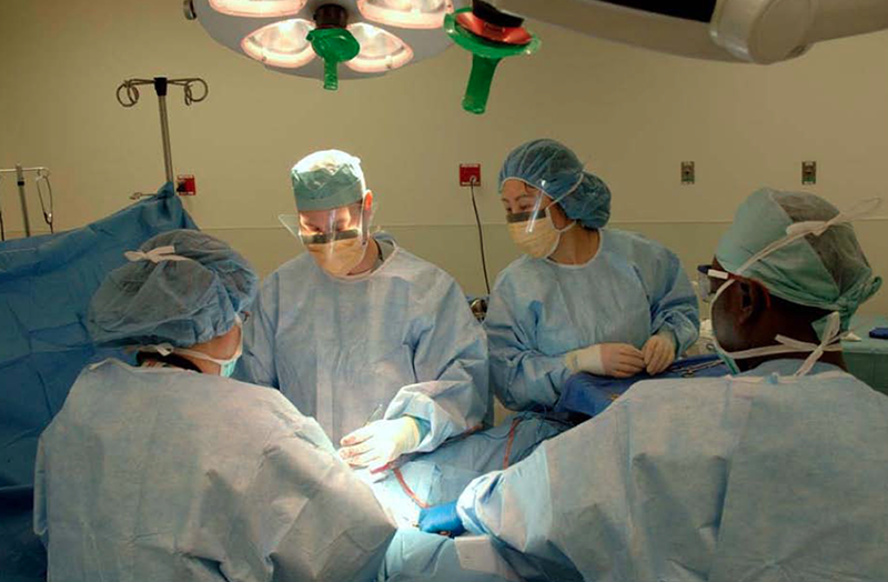
Click image to enlarge.
Figure 1. Surgeons in the Unit remove a symptomatic uterine leiomyoma.
The Unit on Reproductive Endocrinology conducts clinical, translational, and basic science studies of uterine leiomyoma.
Fifty percent of women in the United States develop uterine fibroids during their reproductive years, making the condition the most prevalent reproductive disorder of women. Despite its prevalence, the condition remains poorly understood (1). One prominent feature of uterine fibroids is that the cells within the tumors produce a disordered and excessive extracellular matrix (ECM). In earlier work, we examined the characteristics of the cells that might lead to production of this excessive and fibrotic ECM and obtained evidence that mechanical signaling—a method of cell communication and activation—was attenuated in cells within a fibroid (2). Recently, we examined surgical specimens and immortalized leiomyoma and myometrial cells and indeed found that mechanical signaling was lower in leiomyoma cells than in myometrial cells (3). Additional research has suggested that altered vascular perfusion and angiogenesis may play a critical role in fibroid development (4). Furthermore, given that anti-progestins cause reduction in fibroid size, we analyzed surgical specimens from patients treated with anti-progestins. Anti-progesterone–treated fibroids revealed altered phosphorylation of proteins involved in mechanical signaling. Collectively, the findings suggest that altered mechanical signaling and angiogenesis may contribute to leiomyoma growth (2,4).
Role of BRX (also known as AKAP13) in osmotic stress, inflammation, and development
Our previous studies of the gene BRX (now known as AKAP13), which we cloned, indicated that the gene encoded a large Rho-GEF proto-oncoprotein that is involved in estrogen and glucocorticoid receptor activation. In a recent collaborative study with Tomoshige Kino, using mice haploinsufficient for BRX, we found that BRX was critical to the response of lymphocytes to osmotic stress through a pathway involving the nuclear factor of activated T cells 5 (NFAT5) and the scaffold protein JIP4. In prior work, we examined the consequence(s) of haploinsufficiency for AKAP13 and began analysis of the Cre-Lox AKAP13 tissue–specific deletion in the heart of adult mice. In the past year, our studies further support a role for AKAP13 in cardiac function and deepen our understanding of the in vivo function of the gene.
Publications
- Segars JH, Parrott EC, Nagel JD, Guo XC, Gao X, Birnbaum LS, Pinn VW, Dixon D. Proceedings from the third National Institutes of Health International Congress on advances in uterine leiomyoma research: comprehensive review, conference summary, and future recommendations. Hum Reprod Update 2014;20:309-333.
- Jorge S, Chang S, Barzilai JJ, Leppert P, Segars J. Mechanical signaling in reproductive tissues: mechanisms and importance. Reprod Sci 2014;21:1093-1107.
- Malik M, Britten J, Segars J, Catherino WH. Leiomyoma cells in 3-dimensional cultures demonstrate an attenuated response to fasudil, a Rho-kinase inhibitor, when compared to 2-dimensional cultures. Reprod Sci 2014;21:1126-1138.
- Tal R, Segars JH. The role of angiogenic factors in fibroid pathogenesis: potential implications for future therapy. Hum Reprod Update 2014;20:194-216.
- Levy G, Malik M, Britten J, Gilden M, Segars JH, Catherino WH. Liarozole inhibits transforming growth factor-β3--mediated extracellular matrix formation in human three-dimensional leiomyoma cultures. Fertil Steril 2014;102:272-281.
Collaborators
- Alicia Armstrong, MD, Contraceptive Discovery and Development Branch, NICHD, Rockville, MD
- Paul Driggers, PhD, Uterine Fibroid Tissue Bank, USUHS-NICHD, Bethesda, MD
- Tomoshige Kino, PhD, Program in Reproductive and Adult Endocrinology, NICHD, Bethesda, MD
- Phyllis Leppert, MD, PhD, Duke University, Durham, NC
- Lynnette Nieman, MD, Program in Reproductive and Adult Endocrinology, NICHD, Bethesda, MD
- Pamela G. Robey, PhD, Craniofacial and Skeletal Diseases Branch, NIDCR, Bethesda, MD
- Bradford J. Wood, MD, Center for Interventional Oncology, NIH Clinical Center, Bethesda, MD
Contact
For more information, email segarsj@mail.nih.gov.