


You are here: Home > Section on Cellular Signaling
Signaling and Secretion in Neuroendocrine Cells
- Stanko S. Stojilkovic, PhD, Head, Section on Cellular Signaling
- Silvana A. Andric, PhD, Special Volunteer
- Claudio E. Coddou Alvarez, PhD, Visiting Fellow
- Anne-Marie Heegaard, MD, PhD, Special Volunteer
- Marek Kucka, PhD, Visiting Fellow
- Shuo Li, PhD, Visiting Fellow
- Melanija Tomic, PhD, Staff Scientist
- Zonghe Yan, MD, PhD, Research Fellow
Using multidisciplinary and collaborative approaches, the Section investigates signaling pathways at the cellular and molecular levels, determines the manner in which hormones and neurotransmitters utilize calcium as an intracellular messenger, and characterizes channels involved in electrical activity and calcium signaling in hypothalamic, pituitary and other cell types. The Section investigated cellular signaling cascades and secretion in neuroendocrine cells, with special emphasis on the interactions between plasma membrane electrical events and receptor-controlled pathways. Currently, we are addressing the issues of how the structural features of pituitary channels relate to their functions and how plasma membrane receptors and the intracellular signaling milieu affect channel activity. For this purpose, we characterize both native and recombinant channels and receptors that were cloned from the pituitary gland. We also analyze the relevance of these channels and pathways to calcium-dependent cellular processes.
Expression and structural–functional characterization of pituitary P2X receptor channels
We cloned five ATP-gated P2X receptor channels (P2XRs) from the pituitary gland: P2X2R, P2X3R, P2X4R, P2X6R, and P2X7R. Our ongoing work focuses on the characterization of the roles of these channels in signaling and secretion in secretory pituitary cells and on the structural-functional characterization of recombinant channels. Recently, we showed by quantitative RT-PCR that mRNA transcripts for the P2X4 subunit are the most abundant in rat anterior pituitary tissue and confirmed P2X4R protein expression by Western blot analysis. Single-cell patch-clamp recordings show that extracellular ATP induced an inward depolarizing current in a majority of thyrotropin-releasing hormone-responsive pituitary cells, which resembled the current profile generated by recombinant P2X4R. The channels were activated and desensitized in a dose-dependent manner and deactivated rapidly. Activation of these channels led to stimulation of electrical activity and promotion of voltage-gated and voltage-insensitive calcium influx. In the presence of ivermectin, a specific allosteric modulator of P2X4Rs, there was an approximately fourfold increase in the maximum amplitude of the ATP-induced inward current, accompanied by an increase in the sensitivity of receptors for ATP, slowed deactivation of receptors, and enhanced ATP-induced prolactin release. These results indicate that thyrotropin-releasing hormone-responsive cells, including lactotrophs, express homomeric and/or heteromeric P2X4R, which facilitate calcium influx and hormone secretion (see references 1 and 2).
Mammalian P2X receptors contain ten conserved cysteine residues in their ectodomains, which form five disulfide bonds (SS1-5). In collaboration with Hana Zemkova, we analyzed the relevance of these SS pairs in rat P2X4 receptor function by replacing one or both cysteines by alanine or threonine, expressing receptors in HEK293 cells, and studying their responsiveness to ATP in the absence and presence of ivermectin. Response to ATP was not altered when both cysteines forming the SS3 bond (C132-C159) were replaced by threonines. Replacement of SS1 (C116-C165), SS2 (C126-C149), and SS4 (C217-C227), but not SS5 (C261-C270), cysteine pairs by threonines resulted in decreased sensitivity to ATP and faster deactivation times. The maximum current amplitude was reduced in SS2, SS4, and SS5 double mutants and could be partially rescued by ivermectin in SS2 and SS5 double mutants. This response pattern was also observed in numerous single-residue mutants, but receptor function was not affected when the 217-cysteine was replaced by threonine or arginine or when the 261-cysteine was replaced by alanine. The results, which recently appeared (Rokic et al., Physiol Rev., in press), suggest that the SS1, SS2 and SS4 bonds contribute substantially to the structure of the ligand-binding pocket, while the SS5 bond located towards the transmembrane domain contributes to receptor gating.
Our collaborative work with Pablo Huidobro-Toro focused on identification of the ectodomain site(s) for mercury action at P2X receptors. We generated two chimeras using the full-size P2X2 subunit, termed P2X2a, and a splice variant lacking a 69-residue segment in the C-terminal, termed P2X2b, as donors for intracellular and transmembrane segments and the P2X4 subunit as the donor for ectodomain segment of chimeras. The potentiating effect of mercury on ATP-induced current was preserved in Xenopus oocytes expressing P2X4/2a chimera, but was absent from oocytes expressing P2X4/2b chimera. Site-directed mutagenesis experiments revealed that the Cys-430 residue mediates effects of mercury on the P2X2a receptor activity. Given that mercury could act as an oxidative stress inducer, we also tested whether hydrogen peroxide and the mitochondrial stress inducers myxothiazol and rotenone mimicked mercury effects. The experiments revealed that these compounds potentiated the ATP-evoked P2X2a-receptor and P2X4/2a-receptor currents, but not P2X2b-receptor and P2X2a-C430A– and P2X2a-C430S–mutant currents, whereas the antioxidants dithiothreitol and N-acetylcysteine prevented the hydrogen peroxide potentiation. Alkylation of Cys-430 residue with methylmethane-thiosulfonate also abolished the mercury and hydrogen peroxide potentiation. The results are consistent with the hypothesis that the Cys-430 residue is an intracellular P2X2a receptor redox sensor. The present findings will permit identification of other members of the P2X receptor family that may also have a redox sensor, unraveling the implications of intracellular cysteine residues on receptor physiology. Taken together, our findings highlight that P2X receptors are not only sensors of extracellular ATP and allosteric modulators, but may also respond to intracellular stimuli that vary depending on the metabolic state of the cell (see reference 3).
Dependence of cyclic nucleotide efflux on membrane potential in pituitary cells
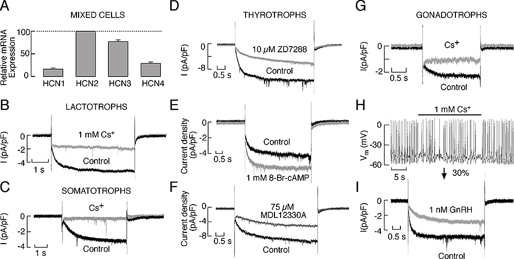
Figure 1. Patterns of spontaneous electrical activity and cyclic nucleotide efflux are determined by sodium gradient.
A-C: Effects of partial substitution of bath sodium with NMDG on the level of hyperpolarization and pattern of electrical activity in GH3 cells (A), lactotrophs (B), and gonadotrophs (C). Inset: the effect of different concentrations of bath sodium on membrane holding current in GH3 cells clamped at -50 mV. D: Effect of partial substitution of extracellular sodium with NMDG on cAMP (top) and cGMP (bottom) efflux in perifused pituitary cells in primary culture. During the treatment, the bath perfusion solution was changed from 145 mM sodium to media containing the sodium concentrations indicated above gray areas, with the residual substituted with NMDG. (click image to enlarge)
In collaboration with Suresh Ambudkar's group, we studied the dependence of multidrug resistance protein–mediated cyclic nucleotide efflux on the background sodium conductance in pituitary cells. These cells fire action potentials and release cyclic nucleotides both spontaneously and in response to agonist stimulation, but the relationship between electrical activity and cyclic nucleotide efflux has not been studied. We showed that a tetrodotoxin-resistant background sodium conductance was critical for firing of action potentials and that the multidrug resistance proteins (MRPs) MRP4 and MRP5 contribute to cyclic nucleotide efflux (see Figure 1). We also showed that abolition of the background sodium conductance in rat pituitary cells by complete or partial replacement of extracellular sodium with organic cations or sucrose induced a rapid and reversible hyperpolarization of cell membranes and inhibition of action potential firing, accompanied by a rapid inhibition of cyclic nucleotide efflux. Valinomycin-induced hyperpolarization of plasma membranes also inhibited cyclic nucleotide efflux, whereas depolarization of cell membranes induced by the inhibition of calcium influx or stimulation of sodium influx by gramicidin was accompanied by a facilitation of cyclic nucleotide efflux. In contrast, inhibition of cyclic nucleotide efflux by probenecid did not affect the background sodium conductance. In human embryonic kidney 293 cells stably transfected with human MRP4 or MRP5, replacement of bath sodium with organic cations also hyperpolarized the cell membranes and inhibited cyclic nucleotide efflux. In these cells, the sodium/hydrogen antiporter monensin did not affect the membrane potential and was largely ineffective in altering cyclic nucleotide efflux. In both pituitary and MRP4- and MRP5-expressing cells, the specific MRP inhibitor 3-[[3-[2-(7-chloroquinolin-2-yl)vinyl]phenyl]-(2-dimethylcarbamoylethylsulfanyl)methylsulfanyl] propionic acid (MK571) inhibited cyclic nucleotide efflux. Our results indicate that the MRP4/5-mediated cyclic nucleotide efflux can be rapidly modulated by membrane potential determined by the background sodium conductance (see references 4 and 5).
Role of calcium in stimulus-secretion coupling and cross-talk between GPCRs
Earlier studies suggested that calcium-controlled exocytosis is mediated by calmodulin-dependent kinase II. To address this hypothesis, we studied the effects of the calmodulin-dependent protein kinase inhibitors isoquinolonesulfonamides KN62 and KN-93 and its inactive analog KN-92 on spontaneous electrical activity, voltage-gated calcium influx, cyclic nucleotide production, and basal prolactin release in pituitary lactotrophs. KN-62 and KN-93 blocked basal prolactin release in a dose- and time-dependent manner. However, a similar effect on basal prolactin release was observed during application of KN-92, which does not inhibit this kinase. KN-93 also inhibited cAMP and cGMP production, but inhibition of prolactin release was independent of the status of cyclic nucleotide production. Single-cell measurements revealed abolition of spontaneous and depolarization-induced electrical activity and calcium transients in KN-92/93–treated cells, with a time course comparable to that observed in secretory studies. The results suggest that caution should be used when interpreting data from studies using isoquinolonesulfonamides to evaluate the role of calmodulin-dependent protein kinases in excitable endocrine cells, because inactive compounds exhibit comparable effects on action potential secretion coupling as active compounds (Popovic et al., Horm Mol Biol Clin Invest. 2010;1:35-42).
In collaboration with Zvi Naor's group, we studied mechanisms of the reciprocal cross-talk between gonadotropin-releasing hormone (GnRH) and prostaglandin receptors. Results of these investigations indicate that GnRH stimulates arachidonic acid release via the calcium-independent phospholipase A2 and not via the more common calcium-dependent cytosolic phospholipase A2. GnRH stimulates COX-1 and COX-2 expression via the protein kinase C-Src/phosphatidylinositol3-kinase/MAPK pathway. COX-2 transcription is apparently mediated by the nuclear factor-B and the CCAAT/enhancer-binding protein sites. The documented inhibition by PGF2 and PGI2 of the autoregulation of GnRH receptor expression is most likely mediated via inhibition of GnRH-stimulated phosphoinositide turnover and not by inhibition of calcium elevation and elevation and activation of MAPK (Naidich et al., Endocrinology 2010;151: 2700-2712).
Additional Funding
- International fellowship award to Dr. Silvana Andric by the Serbian Ministry for Science, Belgrade (451-03-00275) – completed.
- Sabbatical fellowship to Dr. Anne-Marie Heegaard by the Danish Council for Independent Research - Medical Sciences and the Faculty of Pharmaceutical Sciences, University of Copenhagen (2009) – completed.
Publications
- Zemkova H, Kucka M, Li S, Gonzalez-Iglesias AE, Tomic M, Stojilkovic SS. Characterization of purinergic P2X4 receptor channels expressed in anterior pituitary cells. Am J Physiol 2010;298:E644-E651.
- Stojilkovic SS, He M-L, Koshimizu T-A, Balik A, Zemkova H. Signaling by purinergic receptors and channels in the pituitary gland. Mol Cell Endocinol 2010;314:184-191.
- Coddou C, Codocedo JF, Li S, Lillo JG, Acuna-Castillo C, Bull P, Stojilkovic SS, Huidobro-Toro JP. Reactive oxygen species potentiate the P2X2 receptor activity through intracellular Cys430. J Neurosci 2009;29:12284-12291.
- Kucka M, Kretschmannova K, Murano T, Wu C-P, Zemkova H, Ambudkar SV. Stojilkovic SS. Dependence of multidrug resistance protein-mediated cyclic nucleotide efflux on the background sodium conductance. Mol Pharmacol 2010;77:270-279.
- Stojilkovic SS, Tabak J, Bertram R. Ion channels and signaling in the pituitary gland. Endocrine Reviews 2010; 31:doi:10.1210/er.2010-0005.
Collaborators
- Suresh V. Ambudkar, PhD, Center for Cancer Research, NCI, Bethesda, MD
- J. Pablo Huidobro-Toro, PhD, Catholic University, Santiago, Chile
- Zvi Naor, PhD, Tel Aviv University, Israel
- Hana Zemkova, PhD, Institute of Physiology of the Academy of Science of the Czech Republic, Prague, Czech Republic
Contact
For more information, email stankos@helix.nih.gov or visit neuroscience.nih.gov/Faculty/Profile/stanko-stojilkovic.aspx.