You are here: Home > Section on Gamete Development
Cell Cycle Regulation in Oogenesis

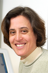
- Mary Lilly, PhD, Head, Section on Gamete Development
- Eva Decotto, PhD, Visiting Fellow
- Remmy Kasili, PhD, Visiting Fellow
- Mary K. Bradford, BS, Undergraduate
- Kuikwon Kim, MS, Technician
- John Csokmay, MD, Clinical Fellow
- Tanveer Akbar, PhD Visiting Fellow
The long-term goal of the laboratory is to understand how the cell-cycle events of meiosis are coordinated with the developmental events of gametogenesis. Chromosome missegregation during female meiosis is the leading cause of miscarriages and birth defects in humans. Recent evidence suggests that many meiotic errors are downstream of defects in oocyte growth and/or the hormonal signaling pathways that drive the differentiation of the oocyte. Thus, an understanding of how meiotic progression and gamete differentiation are coordinated during oogenesis is essential to studies in both reproductive biology and medicine. Currently, we are using the genetically tractable model organism Drosophila melanogaster to examine how meiotic progression is both coordinated with and instructed by the developmental program of the egg.
Meiotic progression and oocyte differentiation in early ovarian cysts
As is observed in mammals and Xenopus, the Drosophila oocyte initiates meiosis within a germline cyst. Drosophila ovarian cysts are produced through a series of four synchronous mitotic divisions during which cytokinesis is incomplete. Soon after the completion of the mitotic divisions, all 16 cells enter premeiotic S phase. However, only the single oocyte remains in meiosis and goes on to produce a functional gamete. The other 15 cells lose their meiotic features, enter the endocycle, and develop as polyploid nurse cells. Throughout much of oogenesis, the oocyte remains faithfully arrested in prophase of meiosis I. Late in oogenesis, the single oocyte undergoes meiotic maturation and proceeds to the first meiotic metaphase. In contrast, the nurse cells transfer their contents to the growing oocyte and undergo apoptosis.
To understand the regulatory inputs that control early meiotic progression, we are working to determine how the oocyte initiates and then maintains the meiotic cycle within the challenging environment of the ovarian cyst. Our studies focus on questions that are relevant to the development of all animal oocytes. What strategies does the oocyte use to protect itself from inappropriate DNA replication? How does the oocyte inhibit mitotic activity prior to meiotic maturation and the full growth and development of the egg? Lastly, how does cell-cycle status within the ovarian cyst influence the differentiation of the oocyte? To answer these questions, we have undertaken studies to determine the basic cell cycle program of the Drosophila ovarian cyst.
The pathways that control entry into the meiotic cycle and early meiotic progression are poorly understood in metazoans. We previously identified a gene, missing oocyte (mio) that regulates nuclear architecture and meiotic progression in early ovarian cysts. In mio mutants, the oocyte enters the meiotic cycle and progresses to pachytene. However, this meiotic state is not maintained, and ultimately the oocyte withdrawals from meiosis, enters the endocycle and becomes polyploid. mio mutants display some of the earliest meiotic defects reported in Drosophila. Moreover, the Mio protein accumulates in the oocyte nucleus in early prophase of meiosis I. Therefore, mio provides an excellent entry point to explore how the unique cell biology of the early ovarian cyst and the establishment of the meiotic program influence the downstream events of oocyte differentiation and meiotic progression.
To better understand the role of mio in oogenesis, we initiated a series of experiments to identify additionally proteins that function in the Mio pathway. From these studies, we have found that Mio physically and genetically interacts with the nucleoporin Seh1, a component of the nuclear pore complex (NPC). The NPC mediates transport of macromolecules between the nucleus and cytoplasm. Recent evidence indicates that structural nucleoporins, the building blocks of the NPC, have a variety of unanticipated cellular functions. We defined an unexpected tissue-specific requirement for the structural nucleoporin Seh1 during Drosophila oogenesis. Seh1 is a component of the Nup107-160 complex, the major structural subcomplex of the NPC. We found that Seh1 associates with the product of the mio gene. Like Mio, the nucleoporin Seh1 has a critical germline function during oogenesis. In both mio and seh1 mutant ovaries, a fraction of oocytes fail to maintain the meiotic cycle and develop as pseudo-nurse cells. Moreover, we found that the accumulation of the Mio protein is greatly diminished in the seh1 mutant background. Surprisingly, our characterization of a seh1 null allele indicates that, while required in the female germline, seh1 is dispensable for the development of somatic tissues. Our work represents the first examination of Seh1 function within the context of a multicellular organism. Our studies demonstrate that Mio is a novel interacting partner of the conserved nucleoporin Seh1 and add to the growing body of evidence that structural nucleoporins can have novel tissue-specific roles. Additionally, our observations provide the framework for future studies on how nuclear pore components influence meiotic progression and maintenance of oocyte identity.
Genomic stability during the premeiotic S phase
The proper execution of premeiotic S phase is essential to both the maintenance of genomic integrity and accurate chromosome segregation during the meiotic divisions. However, the regulation of premeiotic S phase remains poorly defined in metazoa. Cells in the mitotic cycle and the meiotic cycle face a similar challenge. In order to maintain the integrity of the genome, they must replicate their DNA, once and only once, during the S phase. In mitotic cells, this goal is accomplished, at least in part, through the precise regulation of Cdk activity throughout the cell cycle. During the mitotic cycle, Cdk activity inhibits preRC formation. This inhibitory relationship restricts the assemble of preRCs to a short window from late mitosis to G1, when Cdk activity is low, and provides an important mechanism by which mitotic cells prevent DNA rereplication. However, the inhibitory effect of Cdk activity on preRC assembly requires that cells have a strictly defined period of low Cdk activity, prior to S phase, in order to assemble preRCs for the next round of DNA replication. In mammals and yeast, compromising this period of low Cdk activity, by over-expression G1 cyclins, results in decreased replication licensing and genomic instability.
We found that the Cyclin-Dependent Kinase Inhibitor (CKI) Dacapo (Dap) is a key regulator of premeiotic S phase and genomic stability during Drosophila oogenesis. In dap−/− females, ovarian cysts enter the meiotic cycle with high levels of Cyclin E/Cdk2 activity and accumulate DNA damage during the premeiotic S phase. In Drosophila, Cyclin E/Cdk2 activity inhibits the accumulation of the replication-licensing factor Doubleparked/Cdt1 (Dup/Cdt1). Accordingly, we observe that upon entry into the meiotic cycle, Dap promotes the accumulation of Dup/Cdt1. Moreover, mutations in dup/cdt1 dominantly enhance the dap−/− DNA damage phenotype. Importantly, the DNA damage observed in dap−/− ovarian cysts is independent of the DNA double-strand breaks that initiate meiotic recombination. Taken together, our data indicate that the CKI Dap restricts Cyclin E/Cdk2 activity during the early meiotic cycle and is essential for premeiotic S phase and the maintenance of genomic stability in the female germline. Finally, we determined that dap−/− ovarian cysts frequently undergo an extra mitotic division prior to meiotic entry, indicating that Dap influences the timing of the mitotic/meiotic transition.
Publications
- Narbonne-Reveau K, Lilly MA. The Cyclin-dependent kinase inhibitor Dacapo promotes genomic stability during premeiotic S phase. Mol Biol Cell. 2009 20:1960-1969
- Ullah Z, Lee CY, Lilly MA, Depamphilis ML. Developmentally programmed endoreduplication in animals. Cell Cycle 2009 8:1501-1509
Collaborators
- Kim McKim, PhD, Waksman Institute, Rutgers University, Piscataway, NJ
- Jennifer Lippincott-Schwartz, PhD, Cell Biology and Metabolism Program, NICHD, Bethesda, MD
Contact
For more information, email mlilly@helix.nih.gov or visit cbmp.nichd.nih.gov/uccr.


