

You are here: Home > Unit on Microbial Pathogenesis
Host Cell Modulation by the Intracellular Pathogen Legionella pneumophila

- Matthias Machner, PhD, Head, Unit on Microbial Pathogenesis
- Andrew Gaspar, PhD, Postdoctoral Fellow
- M. Ramona Neunuebel, PhD, Postdoctoral Fellow
- Timothy Evans, BSc, MSc, Postbaccalaureate Student
- Yang Chen, BA, Graduate Student
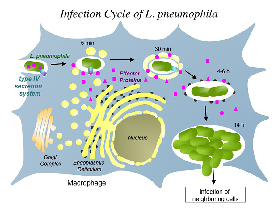
Click image to enlarge.
Figure 1. The infection cycle of L. pneumophila
Many Gram-negative bacteria use type IV secretion systems (T4SSs) to deliver bacterial effector proteins into host cells, where they modulate signaling events to create an environment supportive of bacterial growth. One of those microbes is Legionella pneumophila, the causative agent of a life-threatening pneumonia called Legionnaires' disease. Upon phagocytosis by alveolar macrophages, L. pneumophila delivers a large number of bacterial proteins, or effectors, into the host-cell cytosol through the Dot/Icm T4SS (Figure 1). Some of those effectors help L. pneumophila redirect proteins and membrane material of the infected cell to its Legionella-containing vacuole (LCV), thereby establishing a protective compartment resembling host-cell endoplasmic reticulum (ER). L. pneumophila mutants with a non-functional T4SS are avirulent, underscoring the importance of the translocated effector proteins for L. pneumophila infection. The main focus of our laboratory is to identify and characterize host-pathogen interactions during L. pneumophila infection and to determine their importance for bacterial virulence.
Rab1 recruitment to Legionella-containing vacuoles
One of the trafficking routes from which vesicles are intercepted by L. pneumophila is the early secretory pathway between the ER and the Golgi compartment. Rab1, a key regulator of this pathway, transiently localizes to the LCV, suggesting that L. pneumophila utilizes Rab1 in order to redirect secretory vesicle traffic to its LCV.
We recently discovered that the effector protein SidM is critical for Rab1 exploitation. SidM combines the activity of a GDI displacement factor (GDF) with a guanine nucleotide exchange factor (GEF). SidM mediates dissociation of Rab1 from its chaperone GDP–dissociation inhibitor (GDI) and Rab1 activation by catalyzing exchange of GDP for GTP in Rab1. Bacterial mutants lacking SidM are defective for Rab1 recruitment, indicating that SidM is essential to exploit the pool of GDI–bound Rab1 during infection. SidM is the first known example of a protein shown to combine GDI displacement and nucleotide exchange activity. It remains to be seen if other prokaryotic and eukaryotic proteins exist that share this remarkable ability with SidM.
Reversible AMPylation of Rab1

Click image to enlarge.
Figure 2. The Rab1 cycle on LCV membranes
In addition to its GEF/GDF activity, SidM also catalyzes the covalent attachment of adenosine monophosphate (AMP) to tyrosine-77 of Rab1, a process known as AMPylation. AMPylated Rab1 is locked in the active conformation because it cannot be inactivated by GTPase–activating proteins (GAPs). AMPylated Rab1 is believed to attract cellular cargo vesicles to the LCV without interference from host proteins that would otherwise deactivate Rab1.
Later during infection, the host factor Rab1 eventually disappears from the LCV. The molecular details underlying such removal were unclear. The Legionella effector LepB was believed to inactivate Rab1 by triggering GTP hydrolysis, followed by extraction of Rab1 from the LCV membrane by GDI (which binds only to inactive Rab1). LepB, however, is unable to inactivate Rab1 in its AMPylated form, leaving open the question as to how L. pneumophila can remove AMPylated Rab1 from its LCV. We have now discovered that, in addition to encoding the AMPylase SidM, L. pneumophila produces the effector protein SidD, which catalyzes AMP removal from Rab1, a process known as de-AMPylation (or de-adenylylation). Using purified proteins in in vitro de-AMPylation studies, we found that, once AMP is removed from Rab1 by SidD, de-AMPylated Rab1 is accessible for inactivation by GAP proteins such as L. pneumophila LepB. Using indirect immunofluorescence microscopy, we found that L. pneumophila mutants lacking SidD showed a prolonged colocalization with host cell Rab1, demonstrating that SidD is required for the timely removal of Rab1 from LCVs. A similar phenotype was observed in L. pneumophila mutants deficient in the Rab1-GAP LepB, consistent with the notion that both SidD and LepB collaborate to remove Rab1 from LCVs. The identification of L. pneumophila SidD as Rab1 de-AMPylase not only provides the missing link in a complex chain of Rab1 modulation events during L. pneumophila infection but also represents an intriguing example for how prokaryotic and possibly eukaryotic cells may regulate protein function through reversible AMPylation.
Publications
- Neunuebel MR, Chen Y, Gaspar AH, Backlund Jr. PS, Yergey A, Machner MP. De-AMPylation of the small GTPase Rab1 by the pathogen Legionella pneumophila. Science 2011;333:453-456.
Collaborators
- Peter Backlund, PhD, Biomedical Mass Spectrometry Facility, NICHD, Bethesda, MD
- Alfred L. Yergey, PhD, Biomedical Mass Spectrometry Facility, NICHD, Bethesda, MD
Contact
For more information, email machnerm@mail.nih.gov or visit cbmp.nichd.nih.gov/ump/research.html