

You are here: Home > Section on Genetic Disorders of Drug Metabolism
Molecular Biology, Regulation, and Biochemistry of UDP-Glucuronosyltransferase Isozymes
- Ida S. Owens, PhD, Head, Section on Genetic Disorders of Drug Metabolism
- Nikhil K. Basu, PhD, Staff Scientist
- Sirsendu Jana, PhD, Visiting Fellow
- Amit Raychoudhuri, PhD, Visiting Fellow
- Mousumi Basu, BS, Technician Training Fellow
UDP-glucuronosyltransferase (UGT) isozymes, distributed primarily in the liver, kidney, gastrointestinal tract, and steroid-responsive tissues, carry out the essential function of converting to glucuronides innumerable and frequently encountered structurally diverse lipophilic chemicals that would otherwise be toxic. UGT inactivation of such aromatic-like metabolites and a vast number of dietary, therapeutic, and environmental chemicals reduces the risk of toxicities, mutagenesis and carcinogenesis. Conversion of chemicals to glucuronides inactivates and hastens their excretion from the body, thereby preventing tissue accumulation and toxicities. Neurotoxic bilirubin is the most important endogenous substrate, followed by genotoxic catechol estrogens and dihydrotestosterone at elevated levels. Although the UGT isozyme system prevents bilirubin neurotoxicities in children and inactivates common environmental mutagens and carcinogens, it prematurely converts therapeutics. An understanding of the mechanism of glucuronidation would allow development of methods and strategies for accelerating removal of toxic chemicals while extending the therapeutic benefits of medications that become glucuronidated. In addition to continuing to characterize the individual isozymes that constitute the human UGT chemical defense system, a major research aim of this laboratory has been to understand the basic feature(s)/mechanism(s) underlying the enormous range in substrate selections for UGT isozymes.
Expansion of the phosphorylation requirements of both UGT1A and UGT2B family members
Earlier findings from this laboratory include identification and characterization of: (i) the bilirubin-metabolizing UGT1A1 isozyme; (ii) the novel 215-kb UGT1A locus that encodes 13 independently regulated UGT genes, including the UGT1A1 gene; and (iii) the first known genetic defects in the UGT1A1 gene that lead to Crigler-Najjar diseases in children. We also cloned and identified the primary endogenous substrates for two critical UGT2B isozymes, UGT2B7 and UGT2B15. We continue to study the UGT2B families of isozymes. Characterization of UGT2B7 demonstrated its essential role in detoxifying the depurinating catechol estrogens that are associated with the initiation of breast cancer. Upon cloning of UGT2B15, we demonstrated that it preferentially metabolizes dihydrotestosterone (DHT) and DHT's primary metabolite, 5α-androstane-3α,17β-diol, and that the isozyme is distributed in prostate. Other investigators cloned the companion isozyme UGT2B17 and demonstrated that it also metabolizes the same androgenic compounds, except at 20 to 40-fold higher rates. UGT2B15 was shown to be distributed in prostate luminal cells, while UGT2B17 was found to be distributed in prostate basal cells. UGT2B17 has the same substrate profile as UGT2B15 but with 20 to 40-fold higher activity than UGT2B15. Accordingly, both isozymes participate in DHT homeostasis and thus the prevention of prostate cancer.
Most importantly, we discovered that UGTs require regulated phosphorylation, which can undergo reversible or irreversible downregulation, respectively, by dietary agents or by select kinase inhibitors. Detailed analysis of 5 of 19 human UGTs revealed that regulated phosphorylation occurs at Ser/Thr and/or Tyr residues that are predicted to be PKC and tyrosine kinase sites, respectively, both reversibly inhibited by dietary curcumin. Our model studies with UGT1A7 mutated at PKC sites demonstrated that phosphorylation controls substrate selection, which changes in parallel with changes in pH catalysis optima. Co-immunoprecipitation studies of UGT1A7His and UGT1A10 revealed that PKCepsilon and PKCalpha/delta phosphorylate UGT1A7 and UGT1A10, respectively. Consistent with these observations, treatment of UGT1A7 transfected cells with the PKCepsilon-specific inhibitor peptide or general PKC inhibitors raised 17beta-estradiol catalysis 5- to 11-fold, with a parallel reduction in the level of phospho-serine-432. This novel mechanism involves PKC-mediated phosphorylation of UGT such that phospho-serine/threonine regulates substrate specificity in response to chemical exposures, possibly conferring survival benefit.
While we demonstrated that both PKC and/or tyrosine kinases are, in different manner, involved in the required regulated phosphorylation of UGT isozymes, it was also necessary to established the basis of constitutively active UGTs that depend upon constitutively active PKC support, given that PKC activity can be episodic.
Substrate specificity of UGT2B7 is dictated by differential tyrosine kinase phosphorylation.
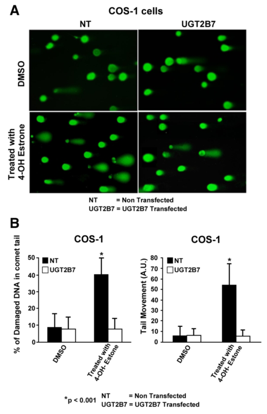
Click image to enlarge.
Figure 1. Effect of UGT2B7 transfection on 4-OHE1–damaged DNA in comet tails
A: Depiction of UGT2B7 protection against 4-hydroxyestrone (4-OHE1)-dependent depurination. B: Quantitation of protection against 4-OHE1-dependent depurination of COS-1 cells. The cells were treated with vehicle or 100 μm 4-OHE1 for 6 h, processed, and stained before evaluation of damaged DNA using Comet Score v1.5 software. The results for 200 cells/group were randomly and cumulatively evaluated from three experiments, using two different parameters as shown: percentage of damaged DNA in tail and tail moment (arbitrary units). Student's t test generated a p value of 0.001.
There is evidence that each UGT isozyme requires distinct phosphorylation owing to different supporting kinase(s). Previously, we confirmed the human UGT2B7 isozyme, which preferentially metabolizes genotoxic 4-OH-catechol estrogens, undergoes tyrosine phosphorylation at two distinct residues following expression in COS-1 cells. UGT2B7 Incorporation of immunoprecipitable [33P]orthophosphate in COS-1 cells and Src in vitro phosphorylation of UGT2B7 expressed in Src-free cells followed by affinity-purification confirmed the phosphorylation. Moreover, our results confirmed that recombinant-expressed UGT2B7 co-localizes with Src kinase, that Y236 and Y438 in UGT2B7 require regulated phosphorylation for its activity, that the isozyme is susceptible to Src kinase inhibitors, and that its phosphorylation by an unidentified tyrosine kinase (TK) in Src-free cells increases its activity 16-fold over that expressed in Src-containing cells. With a highly specific antibody against the Phospho-Y438-2B7, we see lower phospho-density in Src-free cells than in Src-containing cells, which suggests that a more complex phosphorylation pattern exists in Src. Substrate selections by Src-phosphorylated versus the unknown TK in Src-free cells indicate that Src phosphorylated UGT2B7 can essentially metabolize only depurinating 4-OH catechol estrogens, whereas the unidentified TK-phosphorylated UGT2B7 metabolizes these agents at a 10-fold higher rate, as well as metabolizing 17β-estradiol (E2) at a 15-fold higher rate. Hence, Src phosphorylation of UGT2B7 restricts its glucuronidation to the estradiol metabolite rather than allowing glucuronidation of primary estrogen, thereby maintaining the latter's levels for participation in reproduction. While we also confirmed that Src-controlled UGT2B7 prevents toxicity, we showed that UGT2B7 transfected into COS-1 cells efficiently protected against 4-OH-estrone (E1)-mediated depurination. Our results reveal that Src-dependent phosphorylation of UGT2B7 allows metabolism of 4-OHE1, but not of 4-OHE2, in COS-1 cells, while non-Src-phosphorylated UGT2B7 metabolizes both chemicals. Importantly, we determined that UGT2B7 substrate selection is not fixed but varies according to the TK(s) carrying out phosphorylation. Lastly, UGT2B7 transfection of COS-1 cells provided greater than 80 percent protection against 4-OH-estrone-based depurination.
Our attempts to develop a mouse model for the human UGT2B7 to answer related questions under in vivo conditions revealed important differences between human and the mouse UGT2B7 homologues. Determination of UGT2B7 homologue(s) by sequence comparisons identified mouse Ugt-2b34 and Ugt-2b36, which are both distributed in mouse prostate, but not in the mammary gland. In contrast, UGT2B7 is found in the mammary gland but not in prostate. All isozymes are found in liver. Both Ugt2b36 and Ugt2b34 are also distributed in prostate and skin. While Ugt2b34 has exceptionally high activity toward 2-OH-E2/E1, but 73 percent less for 4-OH-E2/E1, Ugt2b36 exhibits nearly exclusive activity toward 4-OH-E2/E1 but is 58 percent less than the maximum seen for Ugt2b34.
An extensive literature search revealed that both human and mouse prostates have both the α and β 17-β-estradiol receptors and that both the stromal cell–distributed α estradiol receptor and the epithelial cell–distributed β estradiol receptor are involved in prostate cancer. In our investigations concerning UGT2b34 and UGT2b36 metabolism of exogenous substrates, we examined toxic chemicals, including designated endocrine disruptors, which are potentially metabolized at the prostate level. To assess protection, we compared UGT2b34 and UGT2b36 in conjunction with prostate UGT2B15 and UGT2B17, which we had identified. For the endocrine disrupters Bisphenol A (BPA) and diethylstilbestrol (DES), we found that Ugt2b34 and Ugt2b36 metabolize both agents. UGT2b34 metabolizes BPA at a high rate with a 4.2-fold preference for BPA, while UGT2b36 metabolizes DES at an even higher rate with a 32-fold preference for DES. Our results demonstrate that, among the active UGTs known to exist in human prostate, only UGT2B15 metabolizes BPA at a very modest rate. BPA leaches from many plastic consumer products, and DES, previously administered to humans for a range of minor to major ailments, was shown to cause imprinting in male fetuses exposed to the agent during gestation, which led to a predisposition to prostate cancer in later adult life.
Dependence of UGT2B15 and UGT2B17 on PKC phosphorylation, with separate regulation by Src kinase phosphorylation
We showed that human prostate–distributed UGT2B15, which metabolizes DHT and its 3α-androstane-5α,17β-diol metabolite, requires regulated phosphorylation in a similar manner to the UGTs we had already analyzed. Reversible downregulation of UGT2B15–transfected COS-1 cells following curcumin-treatment and irreversible inhibition by calphostin-C, bisindolylmaleimide, or Röttlerin treatment versus activation by phorbol 12-myristate 13-acetate indicated that UGT2B15 undergoes PKC phosphorylation. Mutation of three predicted PKC and two tyrosine kinase sites in UGT2B15 caused 70 to 100% and 80 to 90% inactivation, respectively. Gradient SDS-PAGE analysis revealed that anti–UGT-1168 trapped UGT2B15-His–containing co-immunoprecipitates of PKCα in 130-kDa to 140-kDa complexes and larger than 150-kDa complexes. Complexes bound to wild-type UGT2B15-His remained intact during electrophoresis, while UGT2B15-His mutants at phosphorylation sites dissociated to different degrees. PKCα-siRNA treatment inactivated over 50% of COS-1–expressed UGT2B15. In contrast, treatment of UGT2B15–transfected COS-1 cells with the Src-specific activator 1,25 di-hydroxy vitamin D3 enhanced activity, whereas treatment with the Src-specific PP2 inhibitor or Src-siRNA inhibited over 50% of activity. Solubilized UGT2B15-His–transfected Src-free fibroblasts subjected to in vitro [γ33P]ATP-dependent phosphorylation by PKCα and/or Src, affinity-purification, and SDS-gel analysis revealed two-fold greater radiolabeling of 55-kDa to 58-kDa UGT2B15-His by PKCα than by Src, which was additive for combined kinases. Collectively, our evidence indicates that UGT2B15 requires regulated phosphorylation by both PKCα and Src, which is consistent with the complexity of synthesis and metabolism of its major substrate, DHT. Whether basal cells import or synthesize testosterone for transport to luminal cells for reduction to DHT by 5α-steroid reductase-2, comparatively low-activity luminal-cell UGT2B15 undergoes a complex pattern of regulated phosphorylation necessary to maintain homeostatic DHT levels to support occupation of the androgen receptor for prostate-specific functions.
We found that human UGT2B17 expressed in COS-1 cells, which metabolizes DHT and its 5α-androstane-3α,17β-diol (ADT-DIOL) metabolite, requires regulated phosphorylation, as demonstrated by reversible and irreversible inhibition following treatment with curcumin or general PKC inhibitors, respectively. PKCε-siRNA, Src kinase–specific PP2 inhibitor, or Src-siRNA treatment of UGT2B17 expressed in COS-1 cells up-regulated DHT and ADT-DIOL activity, whereas treatment with PKCα-siRNA or PMA in COS-1 cells down-regulated activity. In contrast, treatment of UGT2B17 expressed in SrcYesFyn (SYF)−/− cells with Src–inhibitory agents inhibited UGT2B17. Selective inhibition of DHT glucuronidation over ADT-DIOL for Y99F- and Y237F-UGT2B17 phosphorylation-site mutants expressed in COS-1 cells reached 62% versus 20%, whereas S172A and S422A were null or unaffected, respectively. By contrast, activities for UGT2B17 mutants in SYF-free cells equalized at 50% without evidence of substrate preference, and uniquely PKCε-siRNA treatment inhibited UGT2B17 activity, which confirmed prior up-regulation via Src agents. Western-blot analysis of co-immunoprecipitates of Y237F-UGT2B17-His mutant compared with wild-type UGT2B17-His elicited greater co-migration of PKCα and PKCε in 80kDa to larger than 250-kDa complexe(s). The fact that Y237F-UGT2B17 showed superior PKCε binding indicates that Y99 is a critical residue for tyrosine kinase phosphorylation. Moreover, in vitro studies showed that PKCα, PKCε, and Src individually and additively phosphorylated UGT2B17. For 95% identical prostate-distributed UGT2B17 and UGT2B15, we showed strong PKCε binding at the modified Src Y99 site in UGT2B17 versus UGT2B15, which down-regulated 2B17 activity. Whereas our earlier findings suggest that Src alone phosphorylates UGT2B15 to up-regulate its luminal-cell activity, new evidence indicated that, in contrast, UGT-2B17 is programmed to permit Src down-regulation. A manuscript reporting this study was submitted on 10/1/2012 (Basu et al. Combined positive PKCalpha and negative PKCepsilon and Src effects on regulated phosphorylation of human dihydrotestosterone-metabolizing UGT-2B7 depress its constitutive activity. J Biol Chem).
UGT phosphorylation is requlated via signaling.
As mentioned above, our discovery of rapid, reversible downregulation of human UGTs in LS180 cells following curcumin treatment led to the realization that UGTs require phosphorylation. Having demonstrated that four of five UGTs analyzed to date require PKC–based phosphate signaling for their activity, it was necessary to establish the basis of the constitutively and reliably active PKC to support constitutively active UGTs. Our aim was to determine the relevant kinases and mechanism(s) regulating phosphorylation of constitutive UGTs in LS180 cells and 10 different human UGTcDNA–transfected COS-1 systems. Time- and concentration-dependent inhibition of immunodetectable [33P]orthophosphate in UGTs and PKCε following treatment of LS180 cells with curcumin or the PKC inhibitor calphostin-C suggested that UGT phosphorylation is supported by active PKC(s). Immunofluorescent and co-immunoprecipitation studies with UGT–transfected cells showed co-localization of UGT1A7His and PKCε and of UGT1A10His and PKCα or PKCδ, which was disrupted by PKC inhibitors. Inhibition of UGT activity by PKCε–specific antagonist peptide or by PKCε–targeted destruction with PKCε–specific siRNA and activation of curcumin-downregulated UGTs with typical PKC agonists verified a central role of PKC in glucuronidation. Moreover, in vitro phosphorylation of nascent UGT1A7His by PKCε confirms that the enzyme is a bona fide PKC substrate. Finally, catalase or herbimycin-A inhibition of constitutive or hydrogen peroxide–activated UGTs demonstrated that ROS-related oxidants act as second messengers in maintaining constitutive PKC–dependent signaling, evidently sustaining UGT phosphorylation and activity. Given that cells use signal transduction collectively to detect and respond appropriately to environmental changes, the results described here, combined with our earlier demonstration that specific phospho-groups in UGT1A7 determined substrate selections, suggest that regulated phosphorylation allows adaptations in differential phosphate utilization by UGTs for them to function efficiently.
Tertiary structure of UGTs
To investigate further the requirements of phosphorylation of ER–bound UGTs, we attempted to purify a catalytically active UGT protein for structural analysis. UGT1A7- and UGT2B7-cDNAs, adapted with thrombin/his/myc affinity ligands, not only establish a highly effective UGT–solubilizing system that retains activity but also enabled us to isolate UGT–containing complexes involved in PKC and/or tyrosine kinase signaling pathways, similar to those described for other cellular processes. The complex contained one 58 kDa UGT1A7His and one 110 kDa beta-COP for every two 29 kDa 14-3-3 phospho-serine chaperone proteins. We found that, for PKC–dependent signaling, UGT1A7 associated with RACKepsilon in a 225-kDa adapter complex that included the phosphoserine-dependent 14-3-3 protein. Mutation of UGT1A7His at its 14-3-3 binding sites led to marked instability of solubilized UGT1A7His. Like all UGTs, UGT1A7 has two 14-3-3 binding sites: S162 and T403. Mutations at these sites indicate that 14-3-3 also stabilizes UGT stored at 4°C. Having continued with studies concerning phosphorylation requirements for the different UGT isozymes, with each requiring a different kinase(s) and presumably a different scaffold protein for the signaling complex, it is becoming evident that, to characterize precisely the mechanism by which phosphorylation enables the catalytic process to 'pivot' and instantly change substrate selection, the technology required to achieve purification and reconstitution is not yet available for a membrane-bound target protein such as UGT.
Structural analysis and identification of the common donor-substrate binding site in UGT1A10
Because of the difficulties associated with purifying ER-bound UGTs for structural studies, we carried out homology-based computer modeling to aid analysis. The search uncovered structural homology in Escherichia coli UDP-galactose 4-epimerase. Consistent with predicted similarities involving the common UDP moiety in substrates, Lineweaver-Burke plots showed that UDP-glucose and UDP-hexanol caused competitive inhibition. Among the predicted binding sites N292, K314, K315, and K404 in UGT1A10, we found two informative sets of mutants: the K314R/Q/A/E/G mutants had null activities; that of K404R was 2.7-fold higher than wild type; and K314/E had 50 percent less activity. Scatchard analysis of binding of the affinity ligand 5-azido-uridine-[beta-32P]-diphosphoglucuronic acid to purified UGT1A10-His or UGT1A7-His revealed high- and low-affinity binding sites. UGT1A10-His bound to radio-labeled affinity ligand and, digested with 2-nitro-5-thiocyanobenzoic acid (NTCB), revealed an 11.3-kDa and a 14.3-kDa peptide associated with K314 and K404, respectively. Similar treatment of UGT1A10His-K314A bound to the ligand lacked both peptides; UGT1A10-HisK404R and UGT1A10-HisK404E showed 1.3-fold greater and 50 percent less label in the 14.3-kDa peptide, respectively, compared with UGT1A10-His, without affecting the 11.3-kDa peptide. Scatchard analysis of binding data of the affinity ligand to UGT1A10His-K404R and UGT1A10His-K404E showed a six-fold reduction and a large increase in Kd, respectively. Our results indicate that K314 and K404 are required UDP-glcA binding sites in UGT1A10, that K404 controls activity and high affinity sites, and that K314 and K404 are strictly conserved in 70 aligned UGTs, except for S321—equivalent to K314—in UGT2B15 and UGT2B17 and I321 in the inactive UGT8, which suggests that UGT2B15 and UGT2B17 have suboptimal activity. Our data thus strongly support UDPglcA binding to K314 and K404 in UGT1A10.
Publications
- Basu NK, Kole L, Basu M, Chakraborty K, Mitra PS, Owens IS. The major chemical detoxifying system of UDP-glucuronosyltransferases requires regulated phosphorylation supported by protein kinase C. J Biol Chem 2008;283:23048-23061.
- Banerjee R, Pennington MW, Garza A, Owens IS. Mapping the UDP glucuronic acid binding site in UDP-glucuronosyltransferase-1A10 by homology-based modeling: confirmation with biochemical evidence. Biochemistry 2008;47:7385-7392.
- Mitra PS, Basu NK, Owens IS. Src supports UDP-glucuronosyltransferase-2B7 detoxification of catechol estrogens associated with breast cancer. Biochem Biophys Res Commun 2009;382:651-656.
- Mitra PS, Basu NK, Basu M, Chakraborty S, Saha T, Owens IS. Regulated phosphorylation of a major UDP-glucuronosyltransferase isozyme by tyrosine kinases dictates endogenous substrate selection for detoxification. J Biol Chem 2011;286:1639-1648.
- Chakraborty SK, Basu NK, Jana S, Basu M, Raychoudhuri A, Owens IS. Protein kinase Calpha and Src kinase support human prostate-distributed dihydrotestosterone-metabolizing UDP-glucuronosyltransferase 2B15 activity. J Biol Chem 2012;287:24387-24396.
Collaborators
- Antony McDonagh, PhD, University of California San Francisco, San Francisco, CA
- Masahiko Negishi, PhD, Laboratory of Reproductive and Developmental Toxicology, NIEHS, Research Triangle Park, NC
- Juan Rivera, PhD, Molecular Immunology and Inflammation Branch, NIAMS, Bethesda, MD
- Tapas Saha, PhD, Lombardi Comprehensive Cancer Center, Georgetown University, Washington, D.C.