

You are here: Home > Section on Neuronal Connectivity
Assembly and Function of Chromatic Circuits in Drosophila

- Chi-Hon Lee, MD, PhD, Head, Section on Neuronal Connectivity
- Chun-Yuan Ting, PhD, Staff Scientist
- Thangavel Karuppudurai, PhD, Postdoctoral Fellow
- Yang Lee, PhD, Postdoctoral Fellow
- Tzu-Yang Lin, PhD, Postdoctoral Fellow
- Krishna Melnattur, PhD, Postdoctoral Fellow
- Moyi Li, BA, Biological Laboratory Technician
- Luis Sullivan, BSc, Postbaccalaureate Fellow
Using the Drosophila visual system as a model, we study how neurons form complex yet stereotyped synaptic connections during development and how the assembled neural circuits extract visual attributes, such as color and motion, to guide animal behaviors. Flies' vision is mediated by three types of photoreceptor, R1-6, R7, and R8, each responding to a specific spectrum of light. The three types of photoreceptor project their axons to three distinct layers in the brain, where the cells form synaptic connections with different target neurons. To study visual circuit functions, we combined structural and functional approaches to map visual circuits. Using both light- and electron-microscopy studies, we identified the medulla neurons that are post-synaptic to R7 and R8 photoreceptors. We determined their usage of neurotransmitters and receptors by single-cell transcript profiling. By targeted manipulation of neuronal activity, we establish the roles of these medulla neurons in processing chromatic information. For circuit development, we focus on the formation of synaptic circuits between medulla neurons and R7 and R8 photoreceptors. We use high-resolution imaging techniques and genetic manipulations to delineate the molecular mechanisms that control dendritic patterning and synaptic specificity of the medulla neurons.
Mapping color-vision circuits
Click image to enlarge.
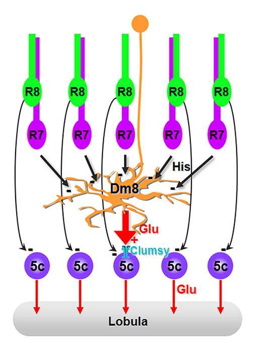
Figure 1. A hard-wired glutamatergic pooling circuit mediates UV preference.
Summary diagram of the R7→Dm8→Tm5c pathway. The amacrine neuron Dm8 receives inhibitory histaminergic inputs from approximately 16 R7 photoreceptors and provides excitatory glutamatergic inputs to one (or at most a few) Tm5c projection neurons. Kainate iGluR receptors expressed in Tm5c are required for receiving glutamatergic inputs from Dm8 and for flies’ UV preference. In addition to the indirect pathway via Dm8, Tm5c neurons also receive direct photoreceptor inputs from R8.
Color vision, which differentiates spectral compositions independent of brightness, provides animals, from insects to primates, great power for object recognition and memory registration and retrieval. In contrast, innate spectral (color) preference, the inborn tendency to be attracted to or repulsed by a specific spectrum of light, is brightness-dependent and reflects unique ecological needs of individual species. Using a combination of genetic, histological, electrophysiological, and behavioral approaches, we study how the visual system processes chromatic information and how this drives various color-driven behaviors. We hypothesize that chromatic information is processed in two discrete stages in Drosophila. First, the transmedulla (Tm) neurons combine multiple photoreceptor channels and relay the information to the lobula, the higher visual center. Second, chromatic information is spatially integrated in the lobula to subserve higher-order color-vision functions, such as color contrast and constancy. Using color preference and color learning assays, we demonstrated that flies innately prefer short wavelengths of light but that they can be trained to select specific wavelengths of light by Pavlovian conditioning, indicating that flies, like honeybees and humans, have true color vision. However, the neural circuits underlying these behaviors differ.
By combining molecular genetics and microscopic techniques, we determined the synaptic circuits of the photoreceptors and their synaptic target neurons in the medulla. The chromatic photoreceptors R7 (UV-sensing) and R8 (blue/green-sensing) provide inputs to a subset of first-order interneurons. The first-order interneurons Tm5a and Tm5b receive direct synaptic inputs from R7s while Tm9, Tm20, and Tm5c receive inputs from R8s. The Tm neurons also receive indirect inputs from R1–6 via L3. The Tm neurons relay spectral information from the medulla to the lobula. In addition to the direct pathways from photoreceptors to Tm neurons, the amacrine neuron Dm8 receives input from several R7s and provides input for Tm5a/b/c. By systematically inactivating and restoring the activity of specific neuronal types and examining behavioral consequences, we determined that the R7→Dm8→Tm5c circuit is specifically required for innate preference for UV light while Tm5a/b/c and Tm20 neurons function redundantly in true color vision.
To relate neural connectivity to function, it is critical to assign components of synaptic machinery to specific connections. To directly probe the usage of neurotransmitters and receptors that determine the polarity and dynamic of signal transmission, we developed a method to profile transcripts in single neurons. We used highly specific promoter–Gal4 constructs to label single types of neurons with GFP; we isolated the GFP–labeled neurons from adult fly brains and profiled their gene expression patterns by RT-PCR. The method enabled us to determine that many of the first-order interneurons in the chromatic circuits express the vesicular glutamate transporter and kainate-type ionotropic glutamate receptors, indicating that interneurons provide and receive fast, sign-conserving glutamate inputs. RNAi–mediated knockdown of the vesicular glutamate transporter in Dm8 amacrine neurons significantly reduced UV preference, suggesting that glutamatergic output of these neurons is critical for its functions. Furthermore, RNAi–mediated knockdown of the kainate receptor Clumsy in Tm5c significantly reduced UV preference. Using a modified GRASP method to probe synaptic connections at the single-cell level, we found that each Dm8 receives about 16 R7 inputs and provides inputs for one, or at most a few, Tm5c neurons in the center of its dendritic field. We conclude that the R7→Dm8→Tm5c connections constitute a hard-wired glutamatergic circuit that pools multiple UV–sensing R7 inputs and relay the information to the lobula to mediate UV preference behavior (Figure 1).
Dendritic development of Drosophila optic lobe neurons
Click image to enlarge.
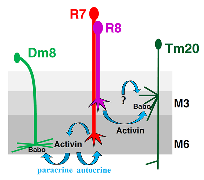
Figure 2. Schematic model illustrating the restriction of target neurons’ dendritic fields by photoreceptor-derived Activin
Activin derived from R8 photoreceptors promotes the dendritic termination of Tm20 neurons and restricts them to their cognate columns. R7–derived Activin plays dual roles: anterograde signaling restricts the dendritic field of the Dm8 neuron, the R7 target, and autocrine signaling restricts R7 growth cones to their retino-topically correct columns.
Our long-term goal is to determine how visual circuits are assembled during development. Most regions of vertebrate and invertebrate brains are organized in columns and layers, which facilitate information processing and propagating. During development, axons and dendrites are routed to specific layers and columns. Spatial coincidence of axons and dendrites affords a key specificity determinant for synaptic pairing. While much knowledge about axonal guidance has been gained over the past years, essentially nothing is known about how target neurons extend dendrites in type-specific patterns to pair with photoreceptors' pre-synaptic terminals. Much of our current understanding of dendrite development came from the studies of the Drosophila Da neurons in the peripheral nervous system, which extend dendrites not to specific targets but to populate (or for tiling) a two-dimensional receptive field. Two key questions concerning dendritic development remain largely unanswered: how CNS neurons establish type-specific dendritic arborization patterns in three-dimension space and how matching occurs in the synaptic partnership between axons and dendrites. Using the Drosophila optic lobe neurons as a model, we aim to determine the molecular mechanisms by which medulla Tm neurons extend dendrites to specific layers and columns to match with photoreceptor presynaptic terminals.
As already stated, we use the Drosophila visual neurons as a model to study dendritic development in the central nervous system. Like the vertebrate cortex and retina, the medulla neuropil in the optic lobe is organized in columns and layers, suggesting that fly medulla neurons and vertebrate cortex neurons confront similar challenges in routing their dendrites to specific layers and columns during development. In addition, the fly visual system has several unique advantages: (i) the medulla neurons extend dendritic arbors in a lattice-like structure, facilitating morphometric analysis; (ii) the presynaptic targets for many medulla neuron types were identified in our anatomical studies; (iii) we developed genetic tools for labeling specific classes of medulla neurons and determining their connectivity in previous studies.
We developed novel techniques to analyze dendritic structures in 3-D and to exploit the unique advantages of this system. First, to image reliably the slender dendrites of medulla neurons, we developed a dual imaging technique that generates isotropic 3-D images of dendrites by combining two confocal image stacks collected in orthogonal directions. Second, we developed an image registration technique that makes use of the regular array structures of the optic lobe to standardize dendritic branching patterns. This, in combination with a series of statistical methods we established, allows us to analyze dendritic patterns in 3-D. Third, we established an imaging technique (GRASP) to detect synaptic contacts at the light-microscopic level.
As a proof of principle, we used the techniques to generate a data set of three types of medulla neurons, Tm2, Tm9, and Tm20. As stated above, Tm9 and Tm20 receive input from R8 photoreceptors in their cognate columns and mediate color vision while Tm2 receives inputs from the achromatic R1–6 via L2 and L4 in its cognate columns and mediates motion detection. Our statistical analyses indicated that: (i) the medulla neurons exhibit stereotypic dendritic arbors but the detailed branching pattern and topology are not conserved in each class; (ii) the synaptic partnerships between axons and dendrites are robust and specific; (iii) the layer-specific routing and polarized extension of dendrites are the two most critical determinants of type-specific dendritic patterns; and (iv) Tm2/9/20 neurons extend dendrites in single medulla columns, consistent with their role in processing retinotopic information. Based on these results, we hypothesize that dendritic development in the optic lobe neurons proceeds in two distinct processes: (i) routing dendrites in type-specific fashion, which, at least in part, serves to maximize the possibility of finding appropriate synaptic partners; (ii) forming the appropriate size of the dendritic field; (iii) matching different sections of dendrites with specific afferents, which likely requires specific interactions between axons and dendrites to ensure synaptic specificity.
By examining a series of candidate mutants, we found that TGF-beta signaling regulates the size of the dendritic field of Tm20 and Dm8, and consequently their synaptic partnership with R8 and R7 photoreceptors, respectively. R7- and R8-derived Activin/TGF-beta selectively restricts the dendritic fields of their respective postsynaptic partners, Dm8 and Tm20, to the size appropriate for their functions. Canonical Activin signaling promotes dendritic termination without affecting dendritic routing direction or layer. Tm20 neurons lacking Activin signaling expand their dendritic fields and aberrantly synapse with neighboring photoreceptors. Thus, afferent-derived Activin regulates the dendritic field size of the photoreceptors’ postsynaptic partners to ensure appropriate synaptic partnership (Figure 2).
Publications
- Wardill TJ, List O, Li X, Dongre S, McCulloch M, Ting C-Y, O'Kane CJ, Tang S, Lee C-H, Hardie RC, Juusola M. Multiple spectral inputs contribute to motion discrimination in the Drosophila visual system. Science 2012;336:925-931.
- Meinertzhagen IA, Lee C-H. The genetic analysis of functional connectomics in Drosophila. Adv Genet 2012;80:99-151.
- Kundu M, Kuzin A, Lin T-Y, Lee C-H, Brody T, Odenwald WF. cis-Regulatory complexity within a large non-coding region in the Drosophila genome. PLoS One 2013;8:e60137.
Collaborators
- Mikko Juusola, PhD, The University of Sheffield, Sheffield, United Kingdom
- Matthew McAuliffe, PhD, Division of Biomedical Imaging Research Services Section, CIT, NIH, Bethesda, MD
- Philip McQueen, PhD, Mathematical and Statistical Computing Laboratory, CIT, NIH, Bethesda, MD
- Ian Meinertzhagen, PhD, DSc, Dalhousie University, Halifax, Canada
- Ward F. Odenwald, PhD, Neural Cell-Fate Determinants Section, NINDS, Bethesda, MD
- Nishith Pandya, BA, Division of Biomedical Imaging Research Services Section, CIT, NIH, Bethesda, MD
- Thomas Pohida, MSEE, Division of Computational Bioscience, CIT, NIH, Bethesda, MD
- Randy Pursley, MSEE, Division of Computational Bioscience, CIT, NIH, Bethesda, MD
- Mark Stopfer, PhD, Program in Developmental Neuroscience, NICHD, Bethesda, MD
- Benjamin White, PhD, Laboratory of Molecular Biology, NIMH, Bethesda, MD