

You are here: Home > Section on Environmental Gene Regulation
Regulatory Small RNAs and Small Proteins

- Gisela Storz, PhD, Head, Section on Environmental Gene Regulation
- Aixia Zhang, PhD, Staff Scientist
- Tamira K. Butler, PhD, Postdoctoral Fellow
- Nikita E. Chavarria, PhD, Postdoctoral Fellow
- Michael D. Dambach, PhD, Postdoctoral Fellow
- Yue Hao, PhD, Postdoctoral Fellow
- Andrew B. Kouse, PhD, Postdoctoral Fellow
- Taylor B. Updegrove, PhD, Postdoctoral Fellow
- Hanbo Wang, BS, Graduate Student
- Jameice DeCoster, BS, Postbaccalaureate Intramural Research Training Award Fellow
- Natasha N. Livingston, BS, Technical Intramural Research Training Award Fellow
Currently, we have two main interests: identification and characterization of small noncoding RNAs and identification and characterization of small proteins of less than 50 amino acids. Both small RNAs and small proteins have been overlooked because they are not detected in biochemical assays and the corresponding genes are poorly annotated and missed in genetic screens. However, mounting evidence suggests that both classes of small molecules play important regulatory roles.
Identification and characterization of small regulatory RNAs
During the past 15 years, we carried out several different systematic screens for small regulatory RNA genes in Escherichia coli. The screens, which have included computational searches for conservation of intergenic regions and direct detection after size selection or co-immunoprecipitation with the RNA–binding protein Hfq, are all applicable to other organisms. Most recently, to further extend our identification of small RNAs, particularly antisense RNAs, we mapped all transcription start sites genomic-wide using deep sequencing (1).
Click image to enlarge.
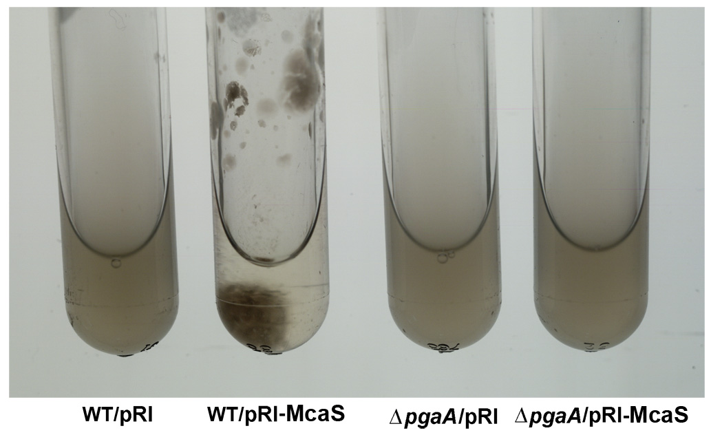
Figure 1. The McaS small RNA induces auto-aggregation of cells.
While cells carrying the vector control (first and third tubes from left) remain suspended, cells overexpressing the McaS small RNA (second tube from left) adhere to the walls and sediment to the bottom of the test tube in the absence of shaking. The phenotype is attributable to McaS activation of the pgaABCD operon required for the synthesis and export of poly-b-1,6-N-acetyl-ᴅ-glucosamine (PGA) and thus is not observed in the pgaA deletion strain (fourth tube from left).
A major focus of the group continues to be the elucidation of the functions of the small RNAs that we and others identified. Early on, we showed that the OxyS RNA, whose expression is induced in response to oxidative stress, acts to repress translation through limited base pairing with target mRNAs. We discovered that the OxyS action is dependent on the Sm-like Hfq protein, which functions as a chaperone to facilitate OxyS RNA base pairing with its target mRNAs. We recently conducted an extensive mutational study of Hfq (2). The analysis revealed that amino acids on three different RNA interaction surfaces—the proximal face, the distal face, and the rim of the doughnut-shaped protein—differentially affect Hfq association with small RNAs and their mRNA targets.
It is now clear that Hfq–binding small RNAs, which act through limited base pairing, are integral to many different stress responses in Escherichia coli. For example, we showed that the Spot 42 RNA, whose levels are highest when glucose is present, plays a broad role in catabolite repression by directly repressing genes involved in central and secondary metabolism, redox balancing, and the consumption of diverse nonpreferred carbon sources. It was previously reported that the transcription factor σE maintains membrane homeostasis, in part by inducing synthesis of proteins involved in membrane repair and of two small regulatory RNAs (sRNAs) that down-regulate synthesis of abundant membrane porins. We recently discovered of a third σE–dependent sRNA, MicL, transcribed from a promoter located within the coding sequence of the cutC gene (3). MicL possesses features typical of Hfq–binding sRNAs but surprisingly targets only a single mRNA, which encodes the outer membrane lipoprotein Lpp, the most abundant protein of the cell. Interestingly, we found that the copper-sensitivity phenotype previously ascribed to inactivation of the cutC gene is, in fact, derived from the loss of MicL and elevated Lpp levels. This observation raises the possibility that other phenotypes currently attributed to protein defects are due to deficiencies in unappreciated regulatory RNAs.
In other recent work, we discovered that the McaS RNA, whose levels are elevated in stationary phase or when glucose is limiting, regulates mRNA targets involved in various aspects of biofilm formation. McaS represses csgD, the transcription regulator of curli biogenesis and activates flhD, the master transcription regulator of flagella synthesis, leading to increased motility. McaS also regulates pgaA, which encodes a porin required for the export of the polysaccharide poly-beta-1,6-N-acetyl-ᴅ-glucosamine (PGA). While we assumed that sRNAs act solely by one mechanism, we discovered to our surprise that McaS acts by two different mechanisms: base-pairing and protein titration (4). McaS base-pairs with the csgD and flhD mRNAs, respectively, resulting in down-regulation and up-regulation of the corresponding cell-surface structures. In contrast, McaS activates pgaA by binding to the global RNA–binding protein CsrA, a negative regulator of pgaA translation. The McaS RNA bears at least two CsrA–binding sequences, and inactivation of these sites compromises CsrA binding, PGA regulation, and biofilm formation. Moreover, ectopic McaS expression leads to induction of two additional CsrA–repressed genes encoding diguanylate cyclases. Thus McaS is a dual-function sRNA with roles in the two major post-transcriptional regulons controlled by the RNA–binding proteins Hfq and CsrA.
In addition to small RNAs that act via limited base pairing, we have been interested in small antisense RNAs that have the potential to form extensive base pairing interactions with their mRNA targets encoded on the opposite strand. We showed that base pairing of GadY, encoded antisense to the gadXW mRNA, results in processing that gives rise to two halves that accumulate to higher levels than the full-length mRNA. We also reported that a large class of antisense RNAs acts to repress the synthesis of small toxic proteins. The lab also made the important discovery that an abundant and broadly conserved small RNA mimics the DNA structure of an open promoter and modulates RNA polymerase activity.
Studies to further characterize other Hfq–binding RNAs and antisense RNAs and to elucidate the roles of small RNAs that act in ways other than base pairing are ongoing.
Identification and characterization of small proteins
In our genome-wide screens for small RNAs, we found that several short RNAs encode small proteins. The correct annotation of the smallest proteins is one of the greatest challenges of genome annotation; perhaps more importantly, the synthesis of only very few small proteins has been confirmed. Although the proteins have largely been missed, the limited numbers of small proteins that have been studied in detail in bacterial and mammalian cells have been shown to have important functions in signaling and cellular defenses (5). We thus established a project to identify and characterize E. coli proteins of fewer than 50 amino acids.
We used sequence conservation and ribosome binding–site models to predict genes encoding small proteins, defined as consisting of 16–50 amino acids, in the intergenic regions of the E. coli genome. We tested expression of the predicted as well as previously annotated small proteins by integrating the sequential peptide affinity tag directly upstream of the stop codon on the chromosome and assaying for synthesis using immunoblot assays. This approach confirmed that 20 previously annotated and 18 newly discovered proteins of 16–50 amino acids are synthesized.
Remarkably, more than half the newly discovered proteins are predicted to be single transmembrane proteins, an observation that prompted us to examine the localization, topology, and membrane insertion of the small proteins. Biochemical fractionation showed that, consistent with the predicted transmembrane helix, the small proteins are generally most abundant in the inner membrane fraction. Examples of both Nin–Cout and Nout–Cin orientations as well as dual topology were found in assays of topology-reporter fusions to representative small transmembrane proteins. Positive residues close to the transmembrane domains are conserved, and mutational analysis of one small protein, YohP, showed that the positive inside rule applies for single transmembrane domain proteins, as has been observed for larger proteins. Also, fractionation analysis of small-protein localization in strains depleted of the Sec or YidC membrane-insertion pathways uncovered differential requirements. Thus, despite their diminutive size, small proteins display considerable diversity in topology, biochemical features, and insertion pathways.
To elucidate the functions of the small proteins, we now are employing many of the approaches the group has used to characterize the functions of small regulatory RNAs. Systematic assays for the accumulation of tagged versions of the proteins showed that many small proteins accumulate under specific growth conditions or after exposure to stress. We also generated and screened bar-coded null mutants and identified small proteins required for resistance to cell-envelope stress and acid shock. In addition, the attached sequential peptide affinity tag is being exploited to identify co-purifying complexes. The combination of these approaches is giving insights into when, where, and how the small proteins act.
We recently showed that expression of a 42–amino acid protein, now denoted MntS (encoded by the small RNA gene formerly known as rybA) is repressed by high levels of manganese through MntR. Overproduction of MntS causes manganese sensitivity, while a lack of MntS perturbs proper manganese-dependent repression of another manganese-regulated gene. Based on these results, we propose that MntS plays a novel role in intracellular manganese trafficking and homeostasis.
We also found that the 49-amino acid inner-membrane protein AcrZ (formerly named YbhT) associates with the AcrAB-TolC multidrug efflux pump, which confers resistance to a wide variety of antibiotics and other compounds in E. coli. Co-purification of AcrZ with AcrB, in the absence of both AcrA and TolC, two-hybrid assays, and suppressor mutations indicate that this interaction occurs through the inner-membrane protein AcrB. The highly conserved acrZ gene is co-regulated with acrAB through induction by the MarA, Rob, and SoxS transcription regulators. Mutants lacking AcrZ are sensitive to many of, but not all, the antibiotics transported by AcrAB-TolC. Such differential antibiotic sensitivity suggests that AcrZ may enhance the ability of the AcrAB–TolC pump to export certain classes of substrates. This work, together with our studies of other small proteins, suggests that many are acting as regulators of larger protein complexes.
Publications
- Thomason MK, Bischler T, Eisenbart SK, Förstner KU, Zhang A, Herbig A, Nieselt K, Sharma CM, Storz G. Global transcriptional start site mapping using dRNA-seq reveals novel antisense RNAs in Escherichia coli. J Bacteriol 2014;E-pub ahead of print.
- Zhang A, Schu DJ, Tjaden BC, Storz G, Gottesman S. Mutations in interaction surfaces differentially impact E. coli Hfq association with small RNAs and their mRNA targets. J Mol Biol 2013;425:3678-3697.
- Guo MS, Updegrove TB, Gogol EB, Shabalina SA, Gross CA, Storz G. MicL, a new σE-dependent sRNA, combats envelope stress by repressing synthesis of Lpp, the major outer membrane lipoprotein. Genes Dev 2014;28:1620-1634.
- Jørgensen MG, Thomason MK, Havelund J, Valentin-Hansen P, Storz G. Dual function of the McaS small RNA in controlling biofilm formation. Genes Dev 2013;27:1132-1145.
- Storz G, Wolf YI, Ramamurthi KS. Small proteins can no longer be ignored. Annu Rev Biochem 2014;83:18.1-18.25.
Collaborators
- Susan Gottesman, PhD, Laboratory of Molecular Biology, NCI, Bethesda, MD
- Carol A. Gross, PhD, University of California San Francisco, San Francisco, CA
- Kay Nieselt, PhD, Universität Tübingen, Tübingen, Germany
- Kumaran S. Ramamurthi, PhD, Laboratory of Molecular Biology, NCI, Bethesda, MD
- Svetlana A. Shabalina, PhD, National Center for Biotechnology Information, NIH, Bethesda, MD
- Cynthia M. Sharma, PhD, Research Centre for Infectious Diseases, Universität Würzburg, Germany
- Poul Valentin-Hansen, PhD, Syddansk Universitet, Odense, Denmark
- Yuri I. Wolf, PhD, National Center for Biotechnology Information, NIH, Bethesda, MD
Contact
For more information, email storz@helix.nih.gov or visit cbmp.nichd.nih.gov/segr.