

You are here: Home > Program in Physical Biology
Program in Physical Biology
Director: Joshua Zimmerberg, MD, PhD
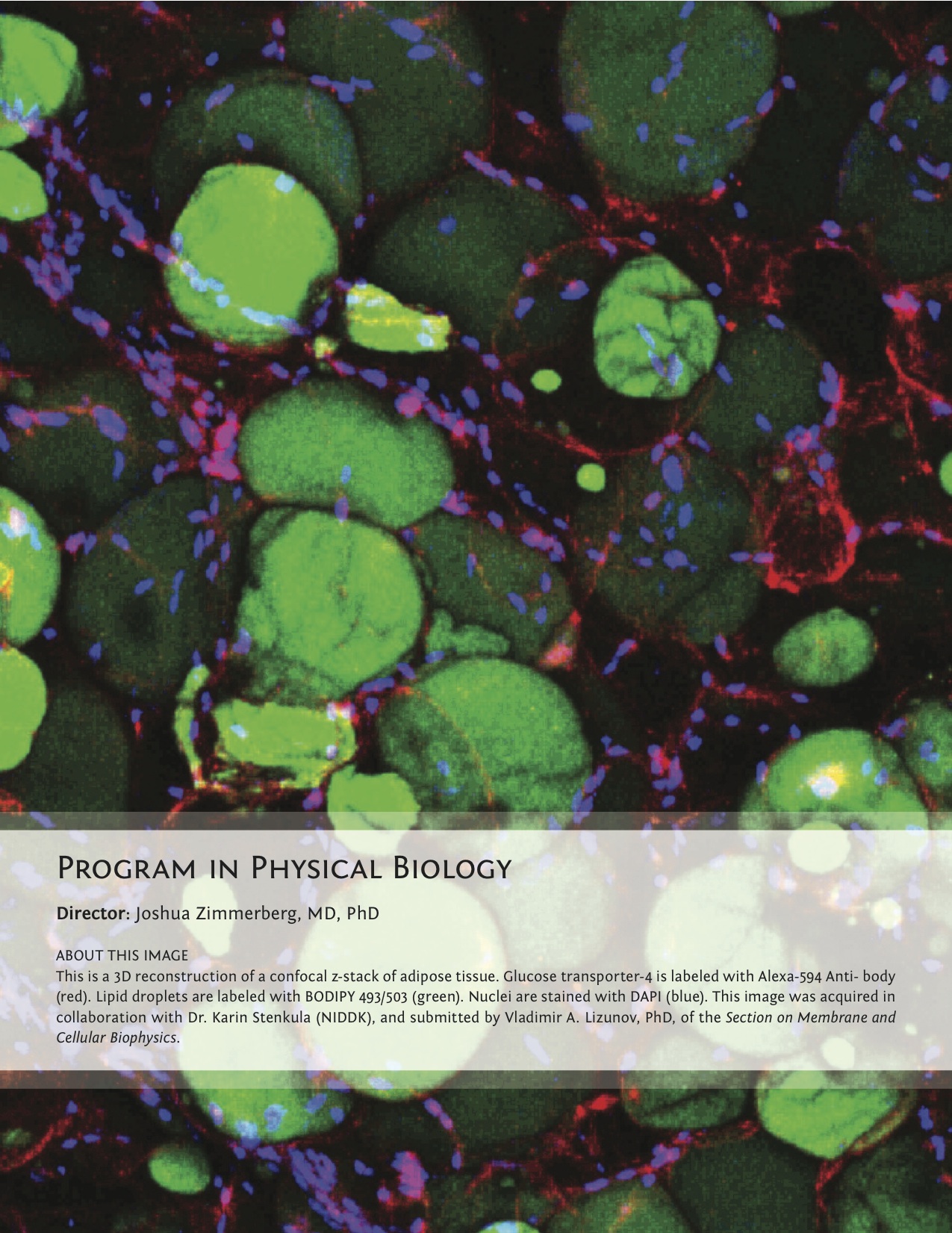
Click image to enlarge.
The Program in Physical Biology (PPB), led by Joshua Zimmerberg, uses systems ranging in complexity from channel-internal surface physics to the network of cytokines and chemokines that determine HIV transmissibility in order to investigate the physicochemical basis of molecular, physiological, and pathological processes and interactions. Research focuses on innovation, on using mathematics to describe biology and pathology, starting with the physical chemistry of surface forces, DNA-protein interactions, microtubular and clathrin polymerization, and on the development of new antiviral drugs and pore-forming antibiotics. PPB scientists use electrophysiology, membrane biochemistry, cell biology, parasitology, immunology, tissue culture, laser micro-dissection, and virology. Diseases of special interest include macular degeneration, diabetes, atherosclerosis, malaria, dengue, HIV, influenza, breast cancer, prostate cancer, fragile sarcolemma muscular dystrophies, and blast-induced traumatic brain injury.
The Section on Molecular Transport, led by Sergey Bezrukov, advances biophysical methods as a tool to understand molecular interactions, notably by studying beta-barrel membrane channel interactions with drugs and cytosolic proteins, as regulated by upstream signaling in the context of human development, disease, and pharmacological intervention. One of the projects aims to unveil the physical mechanisms of regulation of voltage-dependent anion channel (VDAC) of the outer mitochondrial membrane in cell proliferation, reprogrammed cancer metabolism, kinase-regulated cell signaling, and cytoprotection and neurodegeneration, using single-molecule functional approaches. Phosphorylation of VDAC in vitro by either glycogen synthase kinase-3beta or cAMP–dependent protein kinase A increases the on-rate of tubulin binding to VDAC by orders of magnitude. Experiments on HEPG2 human hepatoma cells supported the conjecture that dimeric tubulin and channel phosphorylation regulate VDAC permeability for mitochondrial respiratory substrates. The Section showed that the mechanism of VDAC blockage by tubulin involves tubulin interaction with the membrane as a critical step and that the on-rate of the blockage varies up to 100-fold, depending on the particular lipid composition used for bilayer formation in reconstitution experiments. Immediate physiological implications of these findings include new insights into cellular signaling pathways and cytoskeleton/microtubule activity in health and disease, especially in the case of the highly dynamic microtubule network characteristic of carcinogenesis and cell proliferation.
The Section on Medical Biophysics, led by Robert Bonner, develops new optical technologies to characterize or modify early stressors that drive chronic diseases and for developing effective disease-prevention strategies. Using integrated analysis of multispectral, multimodal clinical retinal imaging, the group maps distributions and dynamics of retinal photochemicals to monitor dysfunction and early disease progression within localized cell clusters. Applying the new methods in clinical studies, the Section seeks to test its novel hypothesis that spectral shifts in retinal irradiance induce retinal photochemical imbalances that drive early age-related and Stargardt's maculopathies and that these could be reduced or prevented by appropriate external filters (e.g., spectral sunglasses). The group also adapted its prior invention of laser capture microdissection into a new low-cost, high throughput device that is being integrated with routine clinical pathology and multiplex molecular analysis. Commercialization of the expression microdissection device is beginning with a focus on enabling improved patient-specific molecular therapy selection. The Section's inventions of new molecular mapping methods are driven by its goal of improving understanding and monitoring of disease states, particularly more readily reversible "preclinical" diseases and their responses to benign, low-cost prevention strategies.
The long-term goal of the Section on Membrane Biology, led by Leonid Chernomordik, is to understand how proteins drive membrane fusion in important cell biology processes. Whereas each kind of protein has its individual personality, membrane lipid bilayers have rather general properties manifested by their resistance to disruption and bending. The analysis of the molecular mechanisms of important and diverse membrane rearrangements will clarify the generality of emerging mechanistic insights and likely bring about new strategies for treating diseases involving cell invasion by enveloped viruses, intracellular trafficking, and intercellular fusion. In recent studies, the Section focused on the membrane-fusion stage of cell entry by dengue virus, the most prevalent mosquito-borne virus. Currently, there are neither vaccines nor effective therapies for dengue infections. As do many enveloped viruses such as influenza and hepatitis C viruses, dengue virus enters the host cell via endocytosis. Fusion between the viral envelope and the endosomal membrane delivers the viral genome into cytosol. Based on their earlier work that identified anionic lipids as a prerequisite for dengue virus fusion, members of the Section designed several novel assays of dengue virus entry and used them to explore mechanisms of virus neutralization by several antibodies. The studies will help in effective vaccine design to prevent dengue disease.
The general goal of the Section on Intercellular Interactions, led by Leonid Margolis, is to understand the mechanisms of pathogenesis and sexual transmission of human pathogens, including the human immunodeficiency virus (HIV), which requires a comprehensive understanding of the mechanisms of pathogenesis in human tissues, where the critical events of the process occurs. Also, such an understanding is a prerequisite for the development of efficient antivirals to contain and/or prevent HIV-1 infection. During the last year, studies focused on: (i) investigating the effects of HIV-1 on the network of cytokines in semen of HIV-1–infected men, which may play an important role in HIV-1 sexual transmission; (ii) developing multi-targeted drugs to contain HIV-I infection and transmission; and (iii) designing new technologies to study immunological mechanisms associated with pathogen transmission and pathogenesis. In particular, the Section found that, in addition to modulating the concentrations of individual cytokines, HIV infection associated with common opportunistic infections alters the entire pattern of the seminal cytokine network. The HIV-1−triggered rearrangement of the seminal cytokine network may contribute to the efficiency of male-to-female HIV-1 transmission. Margolis and his colleagues established that the anti-herpetic drug acyclovir (ACV) inhibits replication of various clinical HIV-1 variants, including ones resistant to commonly approved antivirals. Based on the knowledge of molecular mechanisms of anti-HIV activity of ACV, they developed a way to enhance this activity with ribavirin, another clinically approved drug. Understanding of the basic mechanisms of infection by HIV and other pathogens requires the development of new experimental and diagnostic technologies. This year, the Section's staff reported on one of them, based on nanotechnology. They developed a new method to identify cells that secrete particular cytokines. The research of the Section thus provided new insights into transmission and pathogenesis of HIV-1 and its co-pathogens, leading to new concepts in anti-HIV-1 strategies.
The Section on Cell Biophysics, led by Ralph Nossal, studies cell behavior that can be linked to underlying physical mechanisms, for which the Section develops and applies methodologies based on mathematical and physical principles. The research also uses biochemical and cell-biological techniques. Among current projects are: (i) linking clathrin-coated vesicle biogenesis to physical parameters such as coat rigidity and curvature, energy of coat assembly, receptor diffusion coefficients, and cargo binding energy; (ii) exploring how substrate mechanical properties affect the movement and viability of locomoting eukaryotic cells; (iii) understanding energetic determinants of nucleotide-mediated tubulin associations and how certain small molecules interact with microtubules and thereby act as anti-mitotic agents; and (iv) developing new optical methodologies, based on fluorescence correlation and hyperspectral imaging, to characterize cell function.
The Section on Macromolecular Recognition and Assembly, headed by Donald Rau, focuses on the nature of forces, structure, and dynamics of biologically important assemblies. The group showed that measured forces differ from those predicted by current theories and interpreted the observed forces to indicate the dominant contribution of water-structuring energetics. The observation that interacting macromolecules tenaciously retain their hydration waters unless the surfaces are complementary has profound implications for recognition reactions. To investigate the role of water in binding, the group measures and correlates changes in binding energies and hydration that accompany recognition reactions of biologically important macromolecules, particularly sequence-specific DNA–protein complexes.
The Section on Membrane and Cellular Biophysics, led by Joshua Zimmerberg, studies membranes, viruses, organelles, cells, and tissues in order to understand the molecular organization of cellular membranes, the physico-chemical mechanisms of membrane remodeling, and the molecular anatomy of tissues, which will lead to a better understanding of viral, parasitic, metabolic, developmental, and neoplastic diseases. This past year brought to fruition the following projects: (i) fundamental trafficking mechanisms of glucose homeostasis in muscle; (ii) development of a new hypothesis suggesting that lipid droplets serve as intracellular signaling hubs for insulin resistance caused by a high fat diet; (iii) discovery that secretory vesicles play a role in intracellular calcium homeostasis; (iv) collaboration on the effect of surface clusters on transporter function; (v) development of a new cell line for studying dysferlinopathic musculo-dystrophies (GREG cells) that have already been requested by, and sent around the world to, many investigators of these rare childhood diseases; and (vi) development of a human brain cell culture system, and its use in a newly invented device wherein the pressure waves of a blast (modeled after those found in the Iraq and Afganistan combat theater) are replicated in a microscope stage to determine the cellular and pharmacological mechanisms of blast-induced traumatic brain injury.