


You are here: Home > Program in Physical Biology
Program in Physical Biology
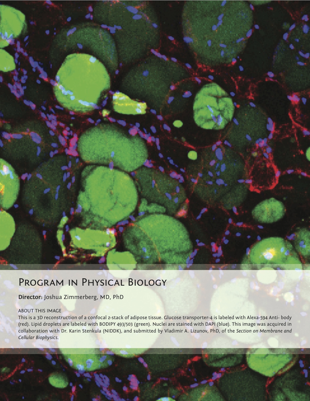
Director: Joshua Zimmerberg, MD, PhD
Click image to enlarge.
The Program on Physical Biology (PPB), led by Joshua Zimmerberg, uses systems ranging in complexity from channel internal-surface physics to HIV pathophysiology in human tissue, to investigate the physicochemical basis of molecular, physiological, and pathological processes and interactions. The PBB develops novel, non-invasive technologies to probe the processes' physical and chemical parameters. The research focuses on the physical chemistry of surface forces, DNA-protein interactions, polymer organic chemistry, membrane biochemistry, pore-forming antibiotics, electrophysiology, cell biology, parasitology, immunology, tissue culture, laser micro-dissection, and virology. Diseases of special interest include macular degeneration, diabetes, malaria, dengue, and HIV. This year, NICHD formed a new program to focus on tissue imaging, comprising the sections of Drs. Basser and Gandjbakhche; V. Adrian Parsegian retired from the NIH. Thus, the PPB is now composed of seven sections.
The Section on Molecular Transport, led by Sergey Bezrukov, focuses on the physical mechanisms underlying regulation of channel-facilitated transport of metabolites and other large solutes across cell and organelle membranes. Among the recent advances is a detailed study of interactions of multi-charged high-affinity toxin inhibitors with the anthrax PA63 pore. The results obtained suggest that, in addition to direct blockage driven by electrostatic interactions, the inhibitors enhance lipid-dependent voltage gating, making the closed states of the channel more favorable. In another project related to the Section's effort to find potent small-molecule inhibitors of pore-forming toxins, the membrane activity of Epsilon toxin from the anaerobic bacterium Clostridium perfringens was studied. The Section showed that Epsilon toxin forms highly asymmetrical conical pores, with the positive charges of the pore shifted to its wider opening. Lastly, the recent progress of the Section in building a comprehensive theory of particle diffusion in confining geometries of transmembrane channels and similar structures should also be lauded.
The Section on Medical Biophysics, led by Robert Bonner, focuses on developing new optical technologies that better characterize or modify the early stressors driving chronic diseases and which could be useful in developing effective disease prevention strategies. The Section continues to develop its inventions of expression microdissection and laser capture microdissection for better integration with multiplex molecular analysis of specific cells and organelles extracted from complex tissue. Through refinements in automated analysis of integrated clinical multispectral, multimodal retinal image sets, the Section also seeks to map the in vivo distribution and dynamics of several naturally occurring photochemicals implicated in health preservation and early disease. In clinical studies, the Section applies its technology approaches to determine how changes in retinal spectral irradiance affect the balance between retinal photochemical pathways that may drive early, more readily reversible disease. The broader goals are to better quantify local molecular imbalances within the retina and to develop reliable means for classifying and quantifying early disease in order to evaluate the effectiveness of strategies to prevent disease progression.
Recent studies performed by the Section on Membrane Biology, led by Leonid Chernomordik, concentrated on the early stages of dengue virus infection. Dengue virus infects up to 100 million people each year and is a leading cause of death among children in some countries. The group identified lipid cofactors required for fusion between dengue virus and the endosomal membrane that delivers viral RNA into cells. The work represents a breakthrough in understanding this important human pathogen and describes first direct assays for the fusion stage of infection. Currently, there are no effective therapies for dengue infections, and new assays will help in developing antivirals, including those targeting newly identified virus-lipid interactions that are found to be essential for dengue virus infection.
The general goal of the Section on Intercellular Interactions, led by Leonid Margolis, is to understand mechanisms of transmission and pathogenesis of human immunodeficiency virus (HIV) in human tissues and to develop efficient anti–HIV-1 preventive and therapeutic strategies. Studies focus on various natural defensive barriers ("gatekeepers") against HIV-1 and are performed in a system of human tissues ex vivo, which recapitulates important aspects of HIV infection in vivo. In particular, an experimental model for male-to-female HIV-1 vaginal transmission was developed to investigate the early barriers in HIV-1 transmission, especially mechanisms of selection for the CCR5-tropic HIV-1 virions that transmit infection through the mucosal barriers. In collaboration with the extra-mural scientists, the Section investigated natural barriers of resistance to HIV infection by using a cohort of uninfected persons in whom risks for infection are high. The working hypotheses pursued by the Section's experiments is that instead of one strong gatekeeper there are many weak barriers whose superimposition is sufficient to protect against X4 HIV-1 infection and potentially select transmitted variants among R5 HIV-1. In general, understanding the "gatekeeping" mechanisms of HIV-1 transmission is critical for the development of effective HIV-1-preventive measures.
The Section on Cell Biophysics, led by Ralph Nossal, aims to understand the physical basis for various basic cell processes. During the past year, the Section 1) used quantitative time-resolved microscopy to show that HIV virions move within cervical mucus in discrete steps probably linked to the relaxation of mechanical stress; 2) investigated conformational fluctuations of clathrin triskelions using atomic force microscopy, showing that the triskelions are flexible in aqueous environments; 3) studied how the presence of high background scattering from an optically dense medium influences the interpretation of data obtained by fluorescence correlation spectroscopy of molecularly crowded solutions; 4) defined the molecular mechanism and binding site of peloruside, the first microtubule-stabilizing drug with a site and mechanism distinct from taxol; and 5) demonstrated proteasome-mediated degradation of tubulin in neural cells exposed to microtubule-depolymerizing chemotherapeutic drugs.
The Section on Macromolecular Recognition and Assembly, headed by Donald Rau, focuses on the nature of forces, structure, and dynamics of biologically important assemblies. The group showed that measured forces differ from those predicted by current theories and interpreted the observed forces to indicate the dominant contribution of water-structuring energetics. The observation that interacting macromolecules tenaciously retain their hydration waters unless the surfaces are complementary has profound implications for recognition reactions. To investigate the role of water in binding, the group measures and correlates changes in binding energies and hydration that accompany recognition reactions of biologically important macromolecules, particularly sequence-specific DNA-protein complexes.
The Section on Membrane and Cellular Biophysics, led by Joshua Zimmerberg, studies membrane mechanics, intracellular molecules, membranes, viruses, organelles, and cells in order to understand viral and parasitic infection, exocytosis, and apoptosis. This past year, research focused on the pathophysiology of two diseases that affect the largest number of people in the world, diabetes and malaria. In particular, the group made three important discoveries. First, insulin causes a differential exposure of the glucose transporter on the surface of adipose cells. Upon delivery to the cell without insulin, there is a cluster of the transporter that does not disperse. With insulin, the transporter is randomly spread across the cell, and it takes time for it to recover by finding partner clusters for internalization. Thus, the key to insulin action involves membrane trafficking. Second, the research showed that there are two more stages to the release of the malaria parasite from red blood cells than originally thought—a swelling stage and a poration stage—which may be targets for therapy. Third, using a novel technique of scanning electron microscopy, new organelles were revealed in the first three-dimensional reconstruction of an infected red cell.