

You are here: Home > Section on Metabolic Regulation
Signal Transduction in Synaptic Transmission and Plasticity

- Kuo-Ping Huang, PhD, Head, Section on Metabolic Regulation
- Freesia L Huang, PhD, Staff Scientist
We investigate the signal transduction mechanisms involved in synaptic transmission and plasticity. Studies of these neural processes are essential to understanding the complex problems related to cognition and behavioral abnormalities. Our current focus is to study genetically modified mice with deletion of the gene encoding neurogranin (Ng), which is specifically expressed in the brain. Ng is a neural-specific protein, which is normally expressed at a high level in subsets of neurons in the forebrain. The protein has been implicated in the regulation of Ca2+/calmodulin (CaM)–dependent cellular processes important for the enhancement of synaptic transmission and plasticity. In humans, mutation of the Ng gene has been linked to behavioral abnormalities and cognitive deficits. In the neuronal soma and dendrites Ng levels are very high, and Ng sequesters apoCaM at basal physiological Ca2+ levels. Upon synaptic stimulation, the influxed Ca2+ displaces Ng from the Ng/apoCaM complex to form Ca2+/CaM and free Ng. The buffering of CaM by Ng serves as a mechanism to regulate neuronal free Ca 2+ and Ca2+/CaM concentrations. The aim of this project is to define the regulatory functions of Ng in neuronal signaling and to design therapeutic approaches to treat cognitive deficits and behavioral disturbances in humans suffering from mutations in Ng.
Stimulation-mediated translocation of calmodulin and neurogranin from soma to dendrites of mouse hippocampal CA1 pyramidal neurons
Interactions of CaM and Ng have been implicated in the enhancement of synaptic plasticity by increasing neurotransmitter-triggered Ca2+ transients. Deletion of the Ng gene in mice causes deficits in learning of hippocampus- and amygdala-dependent behavioral tasks and in high-frequency stimulation (HFS)–induced long-term potentiation (LTP). We found that the concentration of Ng in the hippocampus is approximately twice that of CaM and that the latter protein is largely concentrated in the nucleus and much less in the dendrites. The asymmetrical distribution of CaM in hippocampal neurons is in contrast to that of Ng, which is abundant in the soma and dendrites. Thus, in the distal dendrites, binding of CaM by a relatively higher concentration of Ng renders very little free CaM available for the activation of CaM-dependent enzymes. We explored the possibility that HFS could trigger the mobilization of CaM from soma to dendrites to activate CaMKII and adenylate cyclases for the maintenance of LTP. Tetanic stimulation (a single train of 1 s, 100 Hz) of the Schaffer-collateral fiber of hippocampal slices raised CaM levels in dendrites. We detected HFS–mediated mobilization of CaM in areas surrounding the stimulating electrode within the stratum radiatum but not near a second control electrode that did not deliver HFS. We found that both CaM and Ng associate with the dendritic spines near the stimulating electrode. Translocation of CaM from soma to dendrites was inhibited by the NMDA–receptor antagonist 2-amino-5-phosphonopentanoic acid. We observed no trafficking of CaM in slices from Ng knockout (KO) mice, which exhibited deficits in HFS–mediated LTP. These findings suggest that association of CaM and Ng at the stimulated dendritic spines may enhance synaptic efficacy by increasing the Ca2+ transients as predicted by a "mass-action mechanism." The association of CaM and Ng at the stimulated synapses may also serve as synaptic markers for stimulated dendritic branches.
Calcium-sensitive translocation of calmodulin and neurogranin between soma and dendrites of mouse hippocampal CA1 neurons
Neuronal stimulation that leads to the enhancement of synaptic plasticity requires the activation of Ca2+- and Ca2+/CaM-dependent signaling pathways, which are modulated by Ng through its interaction with CaM. Previous in vitro studies provided ample evidence to support this hypothesis; however, the interaction of Ng and CaM in vivo has yet to be established. We employed acute hippocampal slices to investigate the interaction of these two proteins. Fluorescence immunohistochemical staining of hippocampal slices bathed in Ca2+-containing artificial cerebral spinal fluid (ACSF) revealed that Ng and CaM co-localized in the soma and dendrites of principle neurons. In the CA1 region, the concentration of CaM and Ng in the soma was higher than in the dendrites. Surprisingly, majority of the somatic CaM was sequestered in the nucleus. In contrast, Ng was abundant in the soma as well as in the dendrites. Changing the bathing fluid from a calcium-containing to a calcium-free ACSF induced epileptic activity of the neurons and resulted in subsequent suppression of synaptic transmission. The treatment also caused concomitant redistribution of CaM and Ng from soma to dendrites. Confocal Ca2+ imaging showed that reductions of approximately 15 and 40 nM of intracellular Ca2+ were sufficient to cause half-maximum translocation of Ng and CaM, respectively, from soma to dendrites. Switching the bathing fluid back to Ca2+-containing ACSF restored synaptic transmission and the original compartmentalization of the two proteins. The hippocampal CA1 pyramidal neurons were more responsive to calcium-sensitive translocation than their neighboring CA2 and CA3 neurons. The studies illustrated the unique sensitivity to Ca2+ of the hippocampal CA1 neurons in the mobilization of CaM and Ng between soma and dendrites. Our findings also prove that Ng and CaM respond to changing intracellular calcium in a coordinated manner.
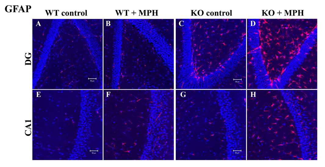
Treatment of neurogranin knockout mice with methylphenidate improves their cognition and behavioral abnormalities.
Click image to enlarge.
Figure 1.
MPH treatment increases GFAP–positive cells in the hilus and stratum radiatum of the CA1 region of the NgKO mouse hippocampus.
Deletion of Ng in mice caused deficits in cognitive functions and HFS-induced LTP in hippocampal slices. Further characterization revealed that the animals exhibited additional behavioral abnormalities, including hyperactivity, impulsivity, and social withdrawal. The behavioral phenotypes likely result from disruption of Ng-regulated signaling. We treated the NgKO mice with methylphenidate (MPH, Ritalin), a psychostimulant drug known to increase extracellular dopamine. Four groups of animals, including control and drug-treated wild-type and NgKO mice, kept in an enriched environment, were injected with MPH (10 mg/kg/day, i.p.) for three weeks and then subjected to behavioral tests. Cognitive functions of the drug-treated NgKO mice improved, as evidenced by a reduction in the latency time to locate the hidden platform in the water maze and an increase in the freezing time after contextual fear-conditioning. MPH also reduced the hyperactivity of NgKO in the open field and increased the immobility time in the forced-swim chamber. The drug-treated mutant mice exhibited improvement in their social interaction with other mice and in recognition of novel mice. The drug treatment, however, had only a marginal effect on the performance of the wild-type mice. Measurement of the HFS-induced (one train of 100 Hz for 1s) LTP in the hippocampal CA1 region in vitro showed a positive effect of the drug on the NgKO hippocampus. At the cellular level, treatment of NgKO with MPH increased glial fibrillary acidic protein (GFAP)–positive astrocytes in the hippocampus, especially prominently in the hilus of dentate gyrus and stratum radiatum of the CA1 region. In the hilus, a large number of astrocytes accumulated at the subgranular layer, which are known to harbor subpopulations of neural stem cells. This structural remodeling may underlie drug-mediated neurobehavioral responses. The results indicate that MPH, a drug commonly used for the treatment of attention-deficit hyperactivity disorder (ADHD), can exert beneficial effects on the NgKO mouse, as it does for the human patients. The studies also suggest that NgKO mice may be useful for the development of new treatment strategies for certain behavioral deficits related to ADHD.
Enhancement of hippocampal synaptic plasticity in neurogranin knockout mice by phorbol ester
One of the most prominent roles of Ng is the enhancement of the NMDA–receptor mediated Ca2+ transients. A rise in intracellular Ca2+ triggers stimulation of Ca2+- and Ca2+/CaM-activated enzymes and their downstream targets to promote synaptic responses and plasticity. Thus, stimulation of downstream signaling components after Ca2+ influx or increase of presynaptic transmitter release may rescue the deficits of NgKO mice. In neurons, phorbol ester is known to enhance transmitter release and facilitate postsynaptic responses by PKC–mediated phosphorylation of AMPA receptors. Short-term treatment (20 min) of hippocampal slices from NgKO mice with the PKC–activating phorbol ester phorbol 12,13-dibutylate (PDBu) or phorbol 12,13-diacetate (PDAc) caused persistent synaptic facilitation, in the hippocampal CA1 region, which lasted for several hours. The phorbol ester–mediated effects were most pronounced among those tissue slices from the dorsal hippocampus that exhibited positive responses in field excitatory postsynaptic potential (fEPSP) and the amplitude of population spike (POPS). In contrast, phorbol ester only enhanced the amplitude of POPS and had no significant effect on the fEPSP of tissue slices from ventral hippocampus. For dorsal hippocampal slices, the initial rate of the phorbol ester–induced stimulation in fEPSP was dose-dependent and was inhibited by the PKC inhibitor chelerythrine but not by the CaMKII inhibitor KN93, the MEK inhibitor U0126, the protein synthesis inhibitor anisomycin, or the NMDA–receptor antagonist APV. Following maximal stimulation by phorbol ester, application of theta-burst electrical stimulation (TBS) caused no additional response. However, prior stimulation with TBS followed by phorbol ester induced additional potentiation of fEPSP, suggesting that the phorbol ester–mediated responses also overlap with those by electrical stimulation, which triggers the activation of postsynaptic NMDA receptors and subsequent stimulation of multiple kinases including PKC. It is intriguing that, for tissue slices from the ventral hippocampus, application of phorbol ester following TBS induced depotentiation. These findings clearly distinguish the physiological responses, to phorbol ester, of the dorsal and from those of the ventral hippocampus. The positive responses of dorsal hippocampus of NgKO mice to phorbol ester–mediated long-term facilitation suggests that treatment of these animals with an analog of the PKC activator (diacylglycerol) may improve the synaptic efficacy of this area, which is thought to be associated with cognitive functions.
Publications
- Huang K-P, Huang FL, Shetty PK. Stimulation-mediated translocation of calmodulin and neurogranin from soma to dendrites of mouse hippocampal CA1 pyramidal neurons. Neuroscience 2011;178:1-12.
- Huang K-P, Huang FL. Calcium-sensitive translocation of calmodulin and neurogranin between soma and dendrites of mouse hippocampal CA1 neurons. ACS Chem Neurosci 2011;2:223-230.
Contact
For more information, email kphuang@helix.nih.gov or visit neuroscience.nih.gov/Faculty/Profile/kuo-ping-huang.aspx