

You are here: Home > Program in Physical Biology
Program in Physical Biology
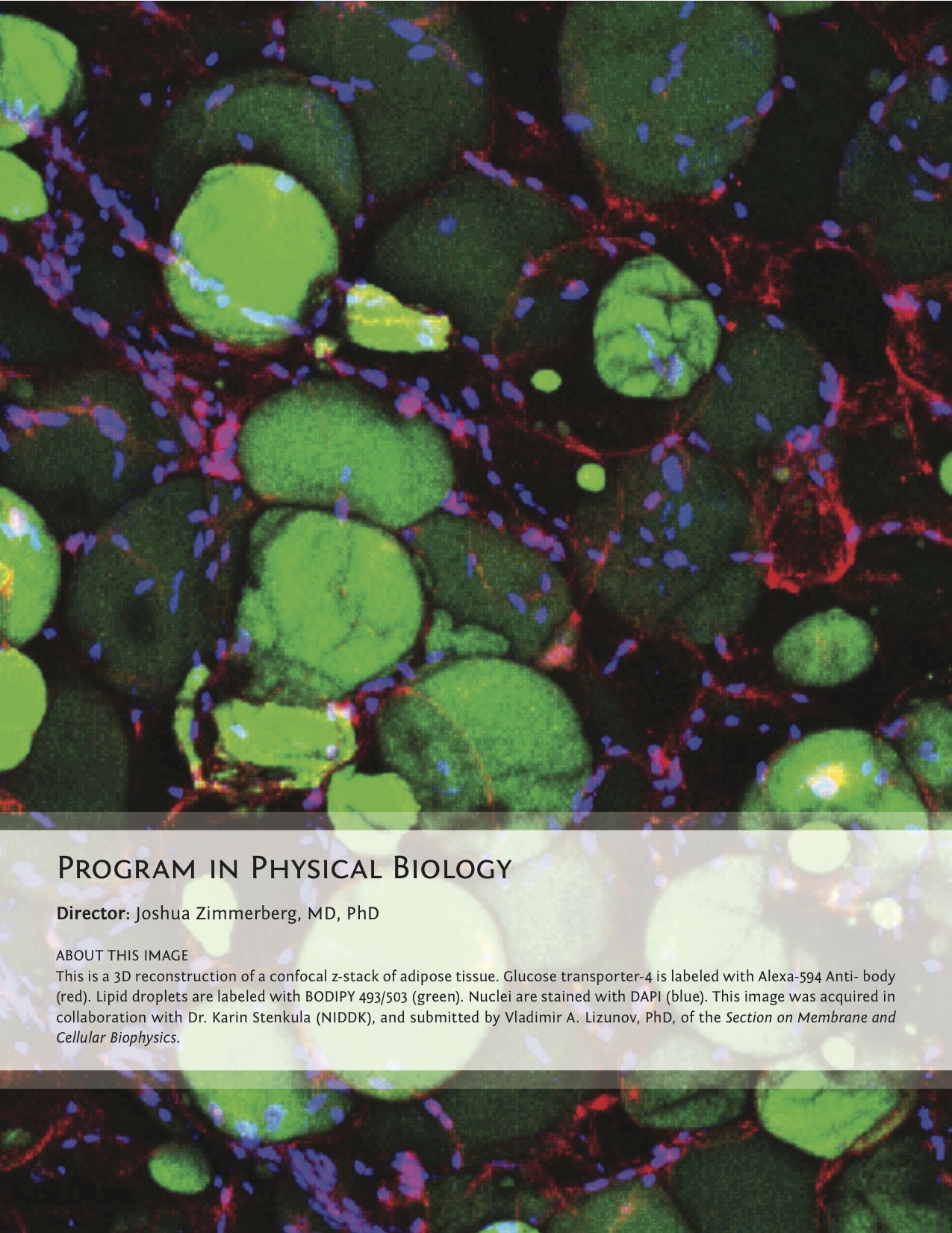
Director: Joshua Zimmerberg, MD, PhD
Click image to enlarge.
Human embryonic development, on which the future child's health depends, is a complex phenomenon within the female starting with egg-spermatozoa fusion. In each individual, a plethora of molecular recognition events mediate the development of an immune system to defend against pathogens, of a musculo-skeletal system to maintain the body, and of flexible networks of molecular expression to manage environmental stress. Traditionally studies on these processes have been divided among biochemistry, cell biology, virology, toxicology, etc. However Nature does not know these artificial divisions, and new understandings are emerging from the crucible that interfaces mathematically-minded physical scientists with biomedical researchers. The Program of Physical Biology, led by Joshua Zimmerberg is a unique scientific body that approaches human development in normal life and pathology as an integral process and that encompass first-class cell biologists, physical chemists, biophysicists, virologists, and immunologists, who not only successfully train post-docs and students within their own fields in work on their own projects but also collaborate widely, building and uniquely promulgating multidisciplinary approaches to the most important biomedical problems in the framework of the NICHD mission.
The Section on Molecular Transport, led by Sergey Bezrukov, advances biophysical methods as a tool to understand molecular interactions, notably by studying, in the context of human development, disease, and pharmacological intervention, the interactions of beta-barrel membrane channel with drugs and cytosolic proteins, as regulated by upstream signaling. One project aims to unveil, using single-molecule functional approaches, the physical mechanisms regulating the voltage-dependent anion channel (VDAC) of the outer mitochondrial membrane in cell proliferation, reprogrammed cancer metabolism, kinase-regulated cell signaling, cytoprotection, and neurodegeneration. The past year's work on the VDAC focused on the effect of lipid composition on the regulation of the interaction between tubulin and VDAC. Surprisingly, changing the composition had effects that were measured in orders of magnitude. Work on toxic pores continued with a study on the interactions of high-affinity cationic blockers with the translocation pores of B. anthracis, C. botulinum, and C. perfringens binary toxins. The physical theory of facilitated transport was also investigated.
The Section on Medical Biophysics, led by Robert Bonner, develops new optical technologies to characterize or modify early stressors that drive chronic diseases and for developing effective disease-prevention strategies. Using integrated analysis of multispectral, multimodal clinical retinal imaging, the group maps the distribution and dynamics of retinal photochemicals to monitor dysfunction and early disease progression within localized cell clusters. Applying the new methods in clinical studies, the Section seeks to test its novel hypothesis that spectral shifts in retinal irradiance induce retinal photochemical imbalances that drive age-related and Stargardt's maculopathies and that these could be reduced or prevented by appropriate external filters (e.g., spectral sunglasses). The group also adapted its prior invention of laser capture microdissection into a new low-cost, high-throughput device that is being integrated with multiplex molecular analysis for application in routine clinical pathology. Commercialization of the expression microdissection device is beginning with a focus on enabling improved patient-specific molecular therapy selection. The Section's inventions of new molecular mapping methods are driven by its goal of improving understanding and monitoring of disease states, particularly more readily reversible "preclinical" diseases and their responses to benign, low-cost prevention strategies.
The long-term goal of the Section on Membrane Biology, led by Leonid Chernomordik, is to understand how proteins drive membrane fusion in important cell-biology processes. Whereas each kind of protein has its individual personality, membrane lipid bilayers have rather general properties manifested by their resistance to disruption and bending. Analysis of molecular mechanisms underlying important and diverse membrane rearrangements will clarify the generality of emerging mechanistic insights and likely bring about new strategies for treating diseases involving cell invasion by enveloped viruses, intracellular trafficking, and intercellular fusion. In recent studies, the Section focused on the cell-to-cell fusion stage during development and regeneration of skeletal muscle and on lipid rearrangements underlying cell entry by cell-penetrating cationic peptides. One surprising discovery was that annexins are the fusogens of myoblast fusion, ending a decades-long search.
The general goal of the Section on Intercellular Interactions, led by Leonid Margolis, is to understand the mechanisms of pathogenesis and sexual transmission of human pathogens, including the human immunodeficiency virus (HIV), which requires comprehensive knowledge of the mechanisms of pathogenesis in human tissues, in particular of the lower female genital tract, where the critical events of this process occur, and which is also a prerequisite for the development of efficient antivirals to contain and/or prevent HIV-1 infection. During the past year, the Section focused on three aims, to: (i) investigate the role of seminal cytokines of HIV-1 infected men in HIV-1 sexual transmission; (ii) develop tissue models in which to study these processes and to use them for testing new multi-targeted drugs to contain HIV-I infection and transmission; and (iii) design modern technologies to study the antigenic composition of individual HIV virions. In particular, the Section found that one of the major seminal cytokines in HIV-1–infected men facilitates virus transmission to a cervico-vaginal ex vivo tissue system. The system reflects many important aspects of the in vivo situation and is now further benchmarked by our studies on the dependence of HIV transmission on menstrual phase. In continuation of their translational research, the Section used the system of cervico-vaginal tissue ex vivo to test new dual-targeting anti-HIV/anti-herpesvirus (HHV) drugs. An understanding the basic mechanisms of infection by HIV and other pathogens also requires development of new experimental and diagnostic technologies. This year, the Section developed a new nanotechnology to characterize the antigenic composition of individual HIV virions, a technology that may reveal which of the HIV virions are the most transmittable/pathogenic, so that the new anti-HIV strategies may specifically target those virions.
The Section on Cell Biophysics, led by Ralph Nossal, studies cell behavior that can be linked to underlying physical mechanisms, for which the Section develops and applies methodologies based on mathematical and physical principles. The research also uses biochemical and cell-biological techniques. Projects currently include: (i) elaborating a physical model to explain the stochastic nature of coated vesicle biogenesis during receptor-mediated endocytosis, focusing on understanding how the size dependence of nanoparticle uptake relies on mesoscopic cell mechanics; (ii) using atomic force spectroscopy to examine the modes by which clathrin lattices can be dissociated; (iii) exploring how substrate mechanical properties affect the response of eukaryotic cells to changes in their environment; and (iv) understanding how certain small molecules interact with microtubules, and thereby act as antimitotic agents, and how microtubule arrays function in mitosis to produce accurate segregation of chromosomes. We also develop new experimental modalities to characterize these and related phenomena. We have a particular interest in the ways cellular activities are coordinated in space and time.
The Section on Macromolecular Recognition and Assembly, headed by Donald Rau, focuses on the nature of forces, structure, and dynamics of biologically important assemblies. The group showed that measured forces differ from those predicted by current theories and interpreted the observed forces to indicate the dominant contribution of water-structuring energetics. The observation that interacting macromolecules tenaciously retain their hydration waters unless the surfaces are complementary has profound implications for recognition reactions. To investigate the role of water in binding, the group measures and correlates changes in binding energies and hydration that accompany recognition reactions of biologically important macromolecules, particularly sequence-specific DNA–protein complexes. This year, projects focused on DNA assembly and compaction, a universal feature of cell biology. The DNA in sperm nuclei is packed densely enough that interhelical spacings can be directly measured by X-ray scattering. During vertebrate spermatogenesis, to achieve compact DNA packaging, chromatin is dramatically reorganized in developing spermatids through replacement of histones with protamines. Given that DNA repair is absent in sperm, dense packaging of DNA in sperm nuclei is considered necessary to protect the DNA against damage by mutagens and reactive oxidizing species (ROS). DNA damage has been shown to correlate with infertility and likely to contribute to miscarriages and birth defects. A current objective is to establish correlations among the DNA packaging density in sperm nuclei, protamine dysfunction, and DNA damage. The observation of the importance of hydration for intermolecular reactions also led the Section to investigate differences in water sequestered by complexes of sequence-specific DNA–binding proteins bound to different DNA sequences, with particular emphasis on correlating binding energy and water incorporated and on the energy necessary to remove hydrating water from complexes. The emphasis on water permits a different approach to recognition reactions than standard practice.
The Section on Membrane and Cellular Biophysics, led by Joshua Zimmerberg, studies membranes, viruses, organelles, cells, and tissues in order to understand the molecular organization of cellular membranes, the physico-chemical mechanisms of membrane remodeling, and the molecular anatomy of tissues, which will lead to deeper insights into viral, parasitic, metabolic, developmental, and neoplastic diseases. Eukaryotic life must create the many shapes and sizes of the system of internal membranes and organelles that inhabit the variety of cells in nature. For cells to secrete signaling macromolecules, express surface transporters, import macromolecular cargo, store energy, and repair damaged plasmalemma, the membranes must remodel. Basic membrane mechanisms require highly regulated and highly organized hierarchies in space and time to allow the organism to thrive despite environmental challenges such as infections by other organisms, unpredictable food supply, and physical trauma. The Section aims to use the expertise and the techniques they perfected over the years to address several biological problems that have in common the underlying regulation or disturbance of protein/lipid interactions. The past year’s projects focused on the discoveries that: intracellular free calcium rises continuously during the egress program of the malaria parasite, rather than spiking; direct chemical imaging of lipid domains in cell membranes reveals a lack of cholesterol domains but confirms sphingomyelin domains; the interactions of the cytoskeleton dictate the dimensions of the clusters of influenza hemagglutinin, just as this molecule itself regulates the components of the cytoskeleton; insulin regulates glucose uptake into fat and muscle by modulating the subcellular distribution of GLUT4 between the cell surface and intracellular compartments in specific insulin-dependent domain dynamics; and GLUT4 trafficking is deficient in cultured adipose cells from insulin-resistant subjects. In a study on the dynamics of protein domains in the catalysis of membrane fission by the GTPase dynamin, a new role—to provide a flexible cage for the reaction to fluctuate—was proposed for these remodeling proteins.