You are here: Home > Section on Developmental Genetics
Heritable Neurodegenerative, Inflammatory, and Autoimmune Disorders

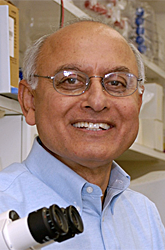
- Anil Mukherjee, MD, PhD, Head, Section on Developmental Genetics
- Zhongjian (Gary) Zhang, MD, PhD, Staff Scientist
- Sondra W. Levin, MD, Adjunct Scientist
- Arjun Saha, PhD, Visiting Fellow
- Chinmoy Sarkar, PhD, Visiting Fellow
- Goutam Chandra, PhD, Visiting Fellow
- Shiyong Peng, PhD, Visiting Fellow
- Eryan Kong, PhD, Visiting Fellow
The Section on Developmental Genetics conducts both laboratory and clinical investigations on the molecular mechanisms of pathogenesis of neurodegenerative, inflammatory, and autoimmune diseases, with a particular emphasis over the past several years on a group of neurodegenerative lysosomal storage disorders (LSDs) mostly affecting children. We focused on understanding the regulation and physiological functions of two genes: 1) palmitoyl-protein thioesterase-1 (Ppt1), mutations of which cause infantile neuronal ceroid lipofuscinosis (INCL), a neurodegenerative LSD, and 2) uteroglobin (UG), which manifests anti-inflammatory and anti-chemotactic properties. We found that ER- and oxidative stresses mediate neuroinflammation in INCL and that omega-3 and that omega-6 fatty acids suppress these stresses, suggesting the fatty acids' therapeutic potential. We also demonstrated that PPT1-deficiency impairs adaptive energy metabolism in neurons, contributing to INCL pathogenesis, and that resveratrol, an antioxidant polyphenol, partially rescues this abnormality. Using UG-knockout (UG-KO) mice, we demonstrated that UG suppresses lung metastasis of B16F10 melanoma cells. Our laboratory and clinical investigations continue to make advances, which are expected to facilitate the development of novel therapies.
Disruption of adaptive energy metabolism contributes to INCL pathogenesis: beneficial effects of resveratrol.
We previously reported that ER and oxidative stresses mediate neuronal apoptosis in both INCL patients and in PPT1-KO mice that mimic INCL. Given that oxidative stress affects mitochondrial function, which is essential for maintaining cellular energy homeostasis, we hypothesized that oxidative stress–mediated disruption of energy metabolism may contribute to INCL pathogenesis. We found that, in cultured INCL fibroblasts and in Ppt1-KO mouse brain, the NAD+/NADH ratio, the levels of phosphorylated-AMPK (p-AMPK), peroxisome proliferator-activated receptor-γ (PPARγ) coactivator-1α (PGC-1α), and silent information regulator-T1 (SIRT1) are markedly down-regulated. This suggested an abnormality in the AMPK/SIRT1/PGC-1α signaling that is required for adaptive energy metabolism. Moreover, we found that, in INCL fibroblasts and in Ppt1-KO mice, phosphorylated-S6K-1 (p-S6K1) levels, which inversely correlate with lifespan, are markedly elevated. Most importantly, dietary resveratrol, an antioxidant polyphenol, elevated the NAD+/NADH ratio and levels of ATP, p-AMPK, PGC-1α and SIRT1, while reducing the p-S6K1 level in both INCL fibroblasts and in Ppt1-KO mice, which showed a modest increase in lifespan. Our results show that oxidative stress–induced disruption of adaptive energy metabolism and increased levels of p-S6K1 contribute to INCL pathogenesis and provide the proof of principle that small molecules such as resveratrol may have therapeutic potential.
Omega-3 and omega-6 fatty acids alleviate apoptosis mediated by oxidative stress in neurons and neuroprogenitor cells from INCL mice.
The nervous system is highly enriched in an enormous variety of complex lipids, among which polyunsaturated fatty acids (PUFAs) predominate. Despite their abundance in the mammalian brain, de novo synthesis of PUFAs such as arachidonic acid (AA) and docosahexanoic acid (DHA) does not occur. Thus, the diet is the sole source of these fatty acids. Epidemiological studies have suggested that increased consumption of omega-3 (n-3) PUFA reduces the risk of neurodegenerative disorders. Neuroprotective effects of PUFA have also been reported, although a clear mechanism has not been defined. PUFA may also have protective effects against oxidative stress, which we previously reported as a mediator of neuronal apoptosis in INCL and in the Ppt1-KO mice. Moreover, emerging evidence indicates that oxygen free radical damage to the brain lipids, carbohydrates, proteins, and DNA may contribute to neuronal cell death, causing neurodegeneration. In the present study, we analyzed cultured PPT1-deficient neurons and neuronal progenitor cells derived from the fetal brain tissues of Ppt1-KO mice. We also explored the effects of PUFA-treatment of these cells, which markedly reduced the level of reactive oxygen species (ROS) and suppressed apoptosis. These results demonstrate for the first time that, while endogenous PUFA levels in the Ppt1-KO mice appear to be similar to or even slightly higher than those of the wild-type littermates, PUFA treatment suppresses oxidative stress and reduces neuronal apoptosis induced by these stresses, raising the possibility that PUFA may therapeutic potential for INCL.
A combination of CystagonTM and Mucomyst® for the treatment of INCL patients: a bench-to-bedside study
INCL is caused by inactivating mutations in the lysosomal Ppt1 gene. Given that PPT1 catalyzes the cleavage of thioester linkages in palmitoylated (S-acylated) proteins, its deficiency impairs degradation or recycling of these lipid-modified proteins by lysosomal proteases. Consequently, accumulation of these modified proteins (ceroids) in lysosomes leads to INCL pathogenesis. Because thioester linkages are susceptible to nucleophilic attack, drugs with this property may have therapeutic potential. Results of our laboratory studies showed that two drugs, phosphocysteamine and N-acetylcysteine, cleave thioester linkages in [14C]palmitoyl-CoA, a model substrate of PPT1, in lymphoblasts and fibroblasts from INCL patients. The drugs also facilitated the depletion of lysosomal ceroids and inhibited apoptosis. The results prompted us to initiate a bench-to-bedside clinical protocol to determine whether a combination of CystagonTM (cysteamine bitartrate) and Mucomyst® (N-acetylcysteine) is beneficial for patients with INCL. Initially, our protocol was approved by NICHD-IRB for treatment of 20 INCL patients; the study has been extended to include 40 patients carrying any two mutations in the Ppt1 gene. So far, we have treated 10 INCL patients; this study is currently recruiting patients and will continue until the approved number of patients are treated.
Lack of an endogenous anti-inflammatory protein in mice enhances metastasis of B16 melanoma cells to the lungs.
Emerging evidence indicates a link between inflammation and cancer metastasis, but the molecular mechanism(s) remains unclear. Uteroglobin (UG), a potent anti-inflammatory protein, is constitutively expressed in the lungs of virtually all mammals. UG-knock-out (UG-KO) mice, which are susceptible to pulmonary inflammation, and B16F10 melanoma cells, which preferentially metastasize to the lungs, provide the components of a model system to determine how inflammation and metastasis are linked. We found that B16F10 cells, injected into UG-KO mice, form markedly more tumor colonies in the lungs than in their wild-type littermates. Remarkably, UG-KO mouse lungs overexpress two calcium-binding proteins, S100A8 and S100A9, whereas B16F10 cells express the receptor for advanced glycation end products (RAGE), which is a known receptor for these proteins. Moreover, S100A8 and S100A9 are potent chemoattractants for RAGE-expressing B16F10 cells, and pretreatment of these cells with a blocking antibody to RAGE suppressed migration and invasion of these cells. Further, in UG-KO mice there is increased gradient of S100A8/S100A9 concentrations from peripheral blood to the lungs, which most likely guides B16F10 cells to the lungs. Moreover, B16F10 cells treated with S100A8 or S100A9 overexpress matrix metalloproteinases, which promote tumor invasion. Taken together, our results suggest that a lack of an endogenous anti-inflammatory protein leads to increased pulmonary colonization of melanoma cells and identify RAGE as a potential anti-metastatic drug target.

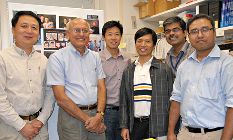
Gary Zhang, Anil Mukherjee, Eryan Kong, Sam Peng, Chinmoy Sarkar, and Goutam Chandra
Additional Funding
- Batten Disease Support and Research Association (BDSRA)
Publications
- Saha A, Lee YC, Zhang Z, Chandra G, Su SB, Mukherjee AB. Lack of an endogenous anti-inflammatory protein in mice enhances colonization of B16F10 melanoma cells in the lungs. J Biol Chem. 2010;285:10822-10831.
- Kim SJ, Zhang Z, Sarkar C, Tsai PC, Lee YC, Dye L, Mukherjee AB. Palmitoyl protein thioesterase-1 deficiency impairs synaptic vesicle recycling at nerve terminals, contributing to neuropathology in humans and mice. J Clin Invest. 2008;118:3075-3086.
- Wei H, Kim S-J, Zhang Z, Tsai PC, Wisniewski KE, Mukherjee AB. ER and oxidative stresses are common mediators of apoptosis in both neurodegenerative and non-neurodegenerative lysosomal storage disorders and are alleviated by chemical chaperones. Hum Mol Genet. 2008;17:469-477.
- Levin SW, Baker E, Gropman A, Quezado ZMN, Miao N, Zhang Z, Jollands A, Di Capua M, Mukherjee AB. Patients with infantile neuronal ceroid lipofuscinosis are susceptible to developing subdural fluid collections. Arch Neurol. 2009;66:1567-1571.
- Zhang Z, Butler JD, Levin SW, Wisniewski K, Brooks SS, Mukherjee AB. A novel approach towards treatment of infantile neuronal ceroid lipofuscinosis, a hereditary progressive encephalopathy of childhood. Nat Med. 2007;7:478-484.
Collaborators
- Eva Baker, MD, PhD, Clinical Center, NIH, Bethesda, MD
- Andrea Gropman, MD, Children's National Medical Center Center for Neuroscience Research (CNR), Washington, D.C.
- Jeeva Munasinghe, PhD, NMR Center, NINDS, Bethesda, MD
- Zenaide Quezado, MD, Departments Anesthesiology and Pain Medicine, Children's National Medical Center, Washington, D.C.
- Yan Xu, PhD, Indiana University Medical School, Indianapolis, IN
Contact
For more information, email mukherja@exchange.nih.gov.


