You are here: Home > Section on Molecular Genetics of Immunity
Gene Regulation in Innate Immunity

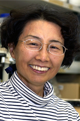
- Keiko Ozato, PhD, Head, Section on Molecular Genetics of Immunity
- Anup Dey, PhD, Biologist
- Tomohiko Kanno, MD, PhD, Staff Scientist
- Natarajan Ayithan, PhD, Visiting Fellow
- Mira Patel, PhD, Visiting Fellow
- Ryusuke Yoshimi, MD, PhD, Supplementary Visiting Fellow
- Naoyuki Sarai, PhD, Visiting Fellow
- Tsung Hsien Chang, PhD, Visiting Fellow
- Monica Gupta, PhD, Visiting Fellow
- Songxiao Xu, PhD, Visiting Fellow
- Walter Huynh, BS, Postbaccalaureate Intramural Research Training Award Fellow
- Daniel Kim, BS, Postbaccalaureate Intramural Research Training Award Fellow
With the goal of understanding gene regulation in the immune system, we study transcription factors and chromatin-binding proteins involved in innate immunity. One of the proteins we work on is the interferon-regulatory factor IRF8, known to direct the development of macrophages and dendritic cells (DC). Macrophages and DC produce proinflammatory cytokines and various chemokines that collectively provide resistance against pathogen infections. IRF8 and other related transcription factors are responsible for inducing cytokines critical for host defense. Their activities are regulated positively and negatively by post-translational modifications. We previously showed that IRF3 and IRF7 are SUMOylated following pathogen stimuli and that SUMOylated IRF3 and IRF7 lose their capacity to activate transcription of type I interferon genes. During the past year, we found that the activities of IRF8 are also regulated by SUMO conjugation. Unlike IRF3 and IRF7, IRF8 was SUMOylated prior to stimulation in macrophages. However, the SUMO moiety was removed following interferon g(IFNg) and toll-like receptor (TLR) stimulation. Interestingly, deSUMOylation coincided with activation of IRF8 function to stimulate cytokine gene transcription. Our results illustrate that proteins of the IRF family are under control of dynamic SUMO conjugation-deconjugation, which affects the pattern of innate immunity.
We also study the role of chromatin in innate immunity and investigate a bromodomain protein Brd4 and the histone variant H3.3. Brd4 binds to acetylated chromatin and recruits transcriptional elongation factor P-TEFb to help enhance transcription. Through its binding to acetyl histones, Brd4 persists on chromatin during mitosis when the bulk of transcription shuts down. For this unusual feature, Brd4 is implicated in the inheritance of transcription across generations of somatic cells. We have begun studying the histone variant H3.3 because of its implied role in epigenetic control: H3.3 is known to be deposited in a replication-independent manner in the actively transcribed regions of the genome. We found that, upon stimulation with IFNβ, Brd4 is recruited to the transcription start sites of all IFN-stimulated genes tested. We now have evidence that this Brd4 recruitment critically controls the subsequent events associated with transcriptional elongation. We further found that IFN-stimulated transcription also causes a remarkable change in chromatin in that it triggers rapid deposition of H3.3. Interestingly, H3.3 incorporation continued long after IFN stimulated transcription ceased. Furthermore shRNA specific for H3.3 significantly reduced IFN-stimulated transcription. Our results indicate that H3.3 is an integral part of transcriptional regulation in innate immunity.
IRF8 acquires transcriptional activator function upon interferon-induced SUMO deconjugation.
Small ubiquitin-like molecules (SUMO), consisting of about 100 amino acids, are conjugated to various substrates through a three-step enzymatic reaction that resembles the ubiquitin conjugation reaction. Similar to ubiquitin conjugation, SUMO conjugation processes are conserved throughout eukaryotes. Many transcription factors are conjugated to SUMO, which, for the most part, causes transcriptional repression. Analogous to reversible ubiquitin conjugation, SUMO conjugation is a reversible process, in that SUMO moieties are removed from the substrates through a set of de-SUMOylating enzymes. In mammalian species, there are seven enzymes involved in catalyzing de-SMOylation. They belong to the SENP family, structurally related to deubiquitinating enzymes. We found that, when RAW cells in the macrophage lineage were stimulated by interferong (IFNg), a large number of nuclear proteins became conjugated to SUMO1. This process was increased when RAW macrophages were further stimulated by TLR ligands. In accordance with IFNg-induced SUMOylation, various types of stress are shown to increase global SUMOylation. Surprisingly, however, IRF8 showed the opposite pattern, as tested by co-immunoprecipitation and immunoblot, in that IRF8 was already conjugated to SUMO prior to stimulation, and levels of SUMOylation markedly decreased after IFNg/TLR stimulation. Using an in vivo SUMO- conjugation assay, we showed that IRF8 is conjugated to SUMO1, SUMO2, and SUMO3 primarily through a lysine (K) residue at 310. This position is within the IAD domain (IRF association domain) and is conserved in humans and mice. We obtained evidence that SUMO conjugation inhibits IRF8's transcriptional activity: 1) an IRF8 mutant K310R gave higher reporter activity for the IL12p40 and IFNβ promoters than wild-type IRF8; 2) in contrast, an IRF8-SUMO fusion protein led to complete inhibition of the reporter activity; 3) although wild-type IRF8 stimulated the development of DCs from bone marrow cells in vitro, the IRF8-SUMO fusion failed to do so. In an effort to delineate mechanisms underlying IRF8 de-SUMOylation, we examined expression of SUMO-deconjugating enzymes and found that SENP1 was induced in RAW macrophages after IFNg/TLR stimulation. Several other SENPs were also induced by the stimulation. Our recent functional analysis using SENP1 shRNA suggests that SENP1 markedly increases IRF8's ability to stimulate Il12p40 and IFNβ promoter activity. These results point to a novel mechanism by which dynamic SUMOylation–de-SUMOylation regulates innate immune responses in macrophages.
The role of Brd4 in transcriptional memory during mitosis
Interaction of Brd4 with acetyl histones persists during mitosis while its interaction with P-TEFb is interrupted during mitosis when most transcription factors, including P-TEFb, are released from chromatin. By detailed ChIP analysis of cells in mitosis, we found that Brd4 localized largely to the transcription start sites (TSS) of genes that are expressed immediately after the completion of mitosis. The genes have been classified as M-G1 genes that include Ran, Kif5b, Topo1,Tgif, Gnb1, Rad21, etc. Genes that are expressed at later times during cell cycle, such as during late G1 S and G2, were largely absent from Brd4. Correlating with Brd4 binding patterns, levels of H3/H4 acetylation were higher in M-G1 genes than in those genes expressed later. While the bulk of RNA polymerase II was released from chromatin during mitosis, some is found at or near the TSS (transcription start site) of most genes expressed in the cells. Furthermore, binding of Brd4 to the TSS dramatically increased at telophase, the last stage of mitosis when post-mitotic transcription starts. The increased binding of Brd4 coincided with a sharp increase in histone H3/H4 acetylation, presumably catalyzed by reassembled histone acetylases. Correlating with this event, the real-time mobility of Brd4 temporarily declined during telophase, as measured by fluorescent recovery after photobleaching, indicating that the affinity of Brd4 for chromatin is increased at telophase. We also noted that P-TEFb is recruited to the TSS of Brd4-bound M-G1 genes at telophase. This in turn coincided with the phosphorylation of the C-terminal domain of RNA polymerase II at serine 2, signifying active transcription of M-G1 genes. To address the importance of the binding of Brd4 to M-G1 genes, we knocked down Brd4 expression by shRNA and found that recruitment of P-TEFb and phosphorylation of RNA polymerase II was markedly reduced in Brd4 knockdown cells. Accordingly, Brd4 knockdown cells exhibited greatly impaired M-G1 gene expression. Our results indicate that Brd4 selectively marks M-G1 genes during mitosis and directs their transcription by mobilizing P-TEFb.
Interferon triggers recruitment of Brd4 and deposition of histone H3.3 in interferon-stimulated genes.
To address the role of Brd4 in innate immunity, we asked whether it is involved in IFN responses. IFNs confer host resistance upon cells by inducing many genes that have anti-viral activity. Using ChIP analyses, we examined recruitment of Brd4 to IFN-stimulated genes (ISGs) in NIH3T3 cells following stimulation with IFNβ. We showed that Brd4 is recruited to the TSS of all ISGs tested, including Ifit1, Mx1, OAS1, and Stat1 immediately following IFN addition, peaking at 6-8 hours, which correlated with ISG mRNA expression. Subsequently, ISG mRNA expression declined and returned to the basal level by 12-24 hours. Along with deminished ISG transcription, Brd4 binding also declined during 12-24 hour, although it never returned to the basal level. A very similar pattern of recruitment was observed for RNA polymerase II and histone acetylation. A series of experiments addressing the functional significance of Brd4 recruitment indicated that Brd4 recruitment is required for subsequent elongation and fine-tuning of ISG transcription.
We also found that the histone variant H3.3 in Brd4 mediated transcription. In an effort to understand the activity of H3.3 in inducible gene expression in the mammalian system, we examined deposition of H3.3 in IFN-stimulated genes in NIH 3T3 cells. Using NIH3T3 cells expressing H3.3 tagged with yellow fluorescent protein (YFP), we tested their deposition in a number of IFN-responsive genes by chromatin immunoprecipitation (ChIP) analysis. Our results showed that H3.3 is deposited rapidly in all IFN-responsive genes tested after stimulation. The most extensive deposition was noted in the 3' end of the coding region of these genes. IFN-responsive genes were transcribed rapidly after stimulation, peaking at around 6 h followed by complete cessation of transcription by 24 h. Surprisingly, H3.3 deposition in these genes continued for at least 24 hours following the completion of transcription. Moreover, H3.3 did not show an increased deposition at the promoter region. The pattern and kinetics of H3.3 deposition was somewhat similar to that of H3K36 trimethylation, as this modification was induced by IFN, and continued even after IFN stimulation. However, H3.3 deposition did not correlate with changes in histone acetylation. The results indicated that H3.3 deposition is coupled with transcriptional activation and has a long-term effect on the chromatin status of IFN-stimulated genes.
Additional Funding
- Trans NIH-FDA Biodefense Program, IATAP Program
Publications
- Kim JY, Ozato K. The sequestosome 1/p62 attenuates cytokine gene expression in activated macrophages by inhibiting IFN regulatory factor 8 and TNF receptor-associated factor 6/NF-kappaB activity. J Immunol. 2009;182:2131-2140.
- Chang TH, Kubota T, Matsuoka M, Jones S, Bradfute SB, Bray M, Ozato K. Ebola Zaire virus blocks type I interferon production by exploiting the host SUMO modification machinery. PLoS Pathogens 2009;6:e1000493.
- Yoshimi R, Chang, TH, Wang H, Atsumi T, Morse H C III, Ozato K. Gene disruption study reveals a non-redundant role for TRIM21/Ro52 in NF-κ B-dependent cytokine expression in fibroblasts. J Immunol. 2009;182:757-7538.
- Kubota T, Matsuoka M, Chaang TH, Bray M, Jones S, Tashiro M, Kato A, Ozato K. Ebolavirus VP35 interacts with the cytoplasmic dynein light chain 8. J Virol. 2009;83:6952-6956.
- Ozato K. PLZF outreach: a finger in interferon pie. Immunity. 2009;30:757-759.
- Zhao B, Takami M, Yamada A, Wang X, Koga T, Hu X, Tamura T, Ozato K, Choi Y, Ivashkiv LB, Takayanagi H, Kamijo R. Interferon regulatory factor-8 regulates bone marrow metabolism by suppressing osteoclastogenesis. Nat Med. 2009;9:1066-1071.
- Dey A, Nishiyama A , Karpova T, McNally J, Ozato K. Brd4 marks select genes on mitotic chromatin and directs post-mitotic transcription. Mol Biol Cell. 2009;23:4899-4909.
- Ozato K, Yoshimi R, Chang TH, Wang H, Atsumi T, Morse HC 3rd. Comment on TRIM21/Ro52 gene disruption. J Immunol. 2009;183:7619-7621.
- Umehara T, Nakamura Y, Jang MK, Nakano K, Tanaka A, Ozato K, Padmanabhan B. Structural basis for acetylated histone H4 recognition by the human BRD2 bromodomain. J Biol Chem. 2010;285:7610-7618.
Collaborators
- Toru Kubota, PhD, Japan National Institute of Infectious Diseases, Tokyo, Japan
- Ben-Zion Levi, PhD, Technion, Israel Institute of Technology, Haifa, Israel
- James McNally, PhD, Laboratory of Receptor Biology and Gene Expression, NCI, Bethesda, MD
- Herbert Morse II, MD, Laboratory of Immunopathology, NIAID, Rockville, MD
- Tomohiko Tamura, MD, PhD, Tokyo University, Tokyo, Japan
Contact
For more information, email ozatok@mail.nih.gov or visit ozatolab.nichd.nih.gov.


