

You are here: Home > Program in Genomics of Differentiation
Program in Genomics of Differentiation
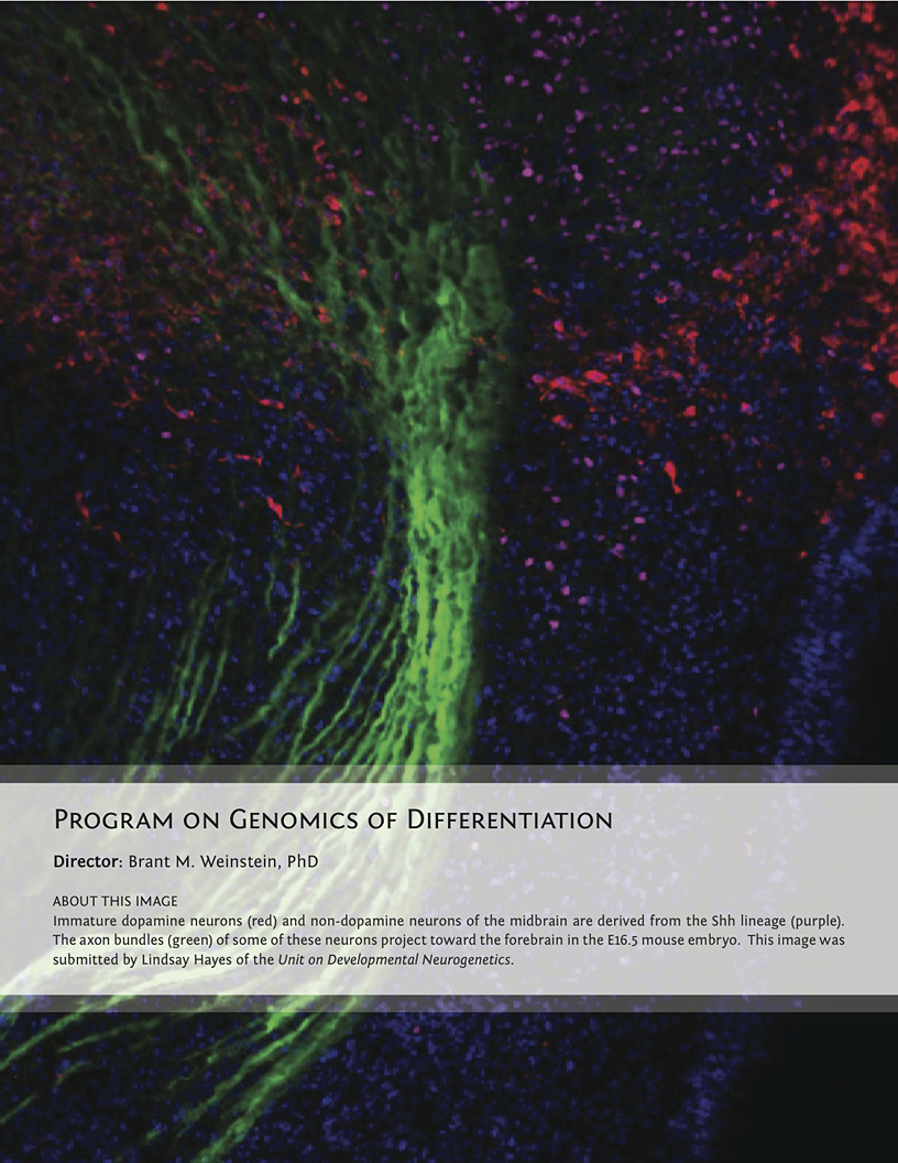
Director: Brant M. Weinstein, PhD
Click image to enlarge.
The Program in Genomics of Differentiation (PGD) is a diverse and highly interactive program in cellular, molecular, and developmental biology research within the Eunice Kennedy Shriver National Institute of Child Health and Human Development (NICHD), Division of Intramural Research (DIR). With 20 principal investigators, the PGD is the largest program in the NICHD DIR, encompassing several research areas, including developmental differentiation and patterning, chromatin dynamics and epigenetics, the immune system, the viral life cycle, DNA replication, gene regulation, and RNA metabolism. Program investigators perform research using a wide variety of models, including viruses, bacteria, mammalian cell culture, yeast, fruit flies, zebrafish, frogs, and mice. Vertebrate models are a major focus of the program. The zebrafish is used as a model for analysis of embryonic development and organogenesis as well as for modeling certain human conditions. Using genetics, genomics, and high-resolution imaging techniques, PGD investigators study cell–cell signaling and cellular behavior in early embryogenesis, formation and morphogenesis of the vascular system, cellular specification in the developing nervous system, and cellular and molecular mechanisms underlying behavior. The mouse provides another important vertebrate model. PGD investigators employ advanced gene-targeting and transgenic technologies to study genes that control mouse development, transcriptional control in the early embryo, the role of Lim-homeobox genes and chromatin-binding proteins, mechanisms of genomic imprinting, regulation of immune cells, the development of the central and peripheral nervous systems, and the behavior of neural stem cells in the adult organism. In addition, the Program generates mouse models of a diverse array of human genetic disorders.
Sohyun Ahn, who heads the Unit on Developmental Neurogenetics, and colleagues continue to investigate the role of Sonic hedgehog (Shh) signaling and its downstream effectors in neurogenesis of the developing and mature brain. One project revealed that the duration of Shh signaling contributes to diversity among dopamine neurons. Combining the power of genetics with other in vivo manipulation techniques, the Unit demonstrated a requirement for Gli3, a major negative regulator of the Shh pathway, in neural progenitors in developing neocortex and in establishing the postnatal neurogenic niche in the subventricular zone of the lateral ventricle. Through comprehensive gene expression–profiling experiments, the Unit also identified additional regulatory signals presented by other cells in the niche, signals that dictate the behavior of neural stem cells. Insights gained from these studies will allow the development of cell type–specific strategies for the recruitment of endogenous neural stem cells during regeneration or disease remediation.
Harold Burgess's Unit on Behavioral Neurogenetics studies the development and function of neural circuits required for behavioral control in larval zebrafish. Brainstem circuits, which control behavior in zebrafish larvae, represent the core of the movement control system in higher vertebrates and are impaired in numerous neurological disorders. The Unit applies computational analysis to high-speed video recordings of larvae challenged with distinct sensory stimuli to determine the function of identified brainstem neurons in transgenic fish. The group generated a library of brain-specific enhancer trap lines and used these to analyze control of sensory responsiveness by environmental context and internal behavioral states such as arousal. The group is currently mapping neuronal circuitry that modulates the startle response to understand how genetic mutations in schizophrenic patients lead to defects in prepulse inhibition.
Mike Cashel's Section on Molecular Regulation discovered two guanine nucleotide analogs (ppGpp and pppGpp) many years ago, when these were called magic spots. The compounds function as a second messenger in bacteria and plants to couple global regulation of gene expression to nutrient availability. This year, the Section's research took three directions. The first was to mutate the ability of the animal kingdom "Mesh" hydrolase to degrade (p)ppApp but not (p)ppGpp in order to use it as a diagnostic tool to match bacterial hydrolase with the reverse specificity. Catalytic-site mutants based on structures are promising. The second was to make a nucleotide test system in E. coli for unusual nucleotides; studies with pGpp were initiated. The third was to exploit the Section's discovery that the omega subunit is part of the (p)ppGpp RNA polymerase binding site. Genetic studies reveal that (p)ppGpp function is not only intertwined with chaperone functions, but that the chaperone activities also appear to be secondary functions of GreA and DksA, which are RNAP accessory proteins involved in initiation, promoter recognition, and elongation arrest.
Ajay Chitnis and colleagues in the Section on Neural Developmental Dynamics are examining how the posterior lateral line system is built in the zebrafish nervous system. The lateral line is a mechanosensory system that consists of sensory organs called neuromasts, which are distributed in a stereotypic pattern over the surface of the zebrafish. Development of hair cells in the lateral line neuromasts is remarkably similar to that of hair cells in the human ear. Furthermore, the mechanisms that guide migration of the lateral line primordium, as it deposits neuromasts under the skin, are remarkably similar to those that determine migration of metastatic cancer cells. The goal of the laboratory is to define the gene-regulatory network that coordinates cell fate and morphogenesis in the lateral line system and to build computational models, based on these studies, to understand how this relatively simple sensory system in zebrafish builds itself.
David Clark and his colleagues in the Section on Chromatin and Gene Expression study the role of chromatin structure in gene activation. Gene activation must occur in the presence of nucleosomes, which are compact structures capable of blocking transcription at every step. To circumvent the chromatin block, eukaryotic cells possess chromatin-remodeling and histone-modifying complexes. Mutations in many of these complexes are strongly associated with cancer. The laboratory used Illumina paired-end sequencing to determine nucleosome positions genome-wide in remodeling mutants of the yeast Saccharomyces cerevisiae and found that the chromatin remodeling complex known as RSC plays a much more important role than the related SWI-SNF complex in organizing yeast chromatin. RSC affects the phasing of nucleosomes relative to the transcription start sites of genes and controls the width of the nucleosome-depleted region that is characteristic of gene promoters. Currently, the laboratory is studying interactions among the various chromatin-remodeling machines to determine their specific roles in chromatin organization.
Robert Crouch, who leads the Section on Formation of RNA, studies RNases H, enzymes that degrade RNA in RNA/DNA hybrids. Failure to degrade RNA in DNA can lead to loss of mitochondrial DNA, detrimental DNA recombination, severe neurological defects, and a general loss of genome integrity. Type I RNase H is structurally and functionally related to an essential RNase H of the HIV-AIDS virus and could be a target for HIV drug therapy. In humans, defective Type II RNase H can result in Aicardi-Goutières syndrome, an encephalopathy that mimics uterine viral infection. In eukaryotes, type II RNase H2 has two distinct activities: removal of single ribonucleotides misincorporated in DNA and of longer stretches of RNA/DNA hybrids, thereby helping to maintain genome stability. Studies on mice and yeast defective in RNase H2 are providing insights into the contribution of this protein to genome integrity.
Igor Dawid, who heads the Section for Developmental Biology, and colleagues study early development in zebrafish and Xenopus. Recent efforts focused on the formation of the neural crest, pineal transcriptome analysis, and the modulation of cell adhesion in the early embryo. In analyzing neural crest development, the group studied the role of the BTB domain protein Kctd15. Kctd15 inhibits neural crest formation, and its action is believed to delimit the neural crest domain in the zebrafish embryo. Kctd15 inhibits transcription factor AP-2, an important neural crest regulator. The mechanism of this inhibition involves the specific binding of Kctd15 to the AP-2 activation domain. SUMO modification of Kctd15 was also studied. This modification is not required for the inhibition of AP-2 by Kctd15, but might have a role in distinct developmental processes. In studying the RNA population of the pineal gland, the group noted a factor, Unc119c, which is differentially expressed in the pineal. Knock-down studies indicate that Unc119c is required in the pineal gland to guide the habenular commissure across the midline in the developing zebrafish brain. Another project showed that the transmembrane E3 ubiquitin ligase March 8 is a modulator of cell-cell adhesion in the early frog and fish embryo.
Mel DePamphilis heads the Section on Eukaryotic DNA Replication, which studies the control of DNA replication and gene expression during mammalian development. The Section's current work focuses on three questions: how genome duplication is restricted to once per cell division in proliferating cells; how these mechanisms can be circumvented in order to selectively kill cancer cells; and how the mechanisms are circumvented during normal mammalian development to allow some cells to differentiate into viable, nonproliferating, polyploid cells. Recently, members of the Section discovered that one of the proteins (Geminin) involved in preventing DNA from re-replicating prior to mitosis is essential to prevent DNA re-replication and apoptosis in cells derived from human cancers, but not in cells derived from normal tissues. They are pursuing this discovery by investigating the effects of Geminin ablation in mice, by using high-throughput screening to identify all genes essential for restricting genome duplication in cancer cells to once per cell division, and by identifying small molecules that selectively target these genes. Such molecules would be useful in cancer chemotherapy as well as in research into the regulation of cell proliferation. To understand how normal cells escape the restrictions on genome duplication, the Section is investigating how trophoblast stem cells exit their mitotic cell cycle and differentiate into the polyploid giant cells essential for both implantation and placentation.
A major effort of the Human Genetics Section, led by Bruce Howard, focuses on epigenome structure, including higher-order chromatin architecture and DNA methylation patterns. The group pays special attention to how defects in the maintenance of epigenetic structures (or failures in programmed transitions, especially in the perinatal period) underlie common developmental disorders and age-related diseases. The group develops and implements bioinformatic tools to facilitate human genome annotation as well as pattern-recognition/-comparison searches of next-generation sequencing data. Together, such unbiased genomics approaches will be invaluable for identifying new features of developmental and age-related of epigenome remodeling.
Judith Kassis, who heads the Section on Gene Expression, studies the mechanism of gene silencing by the Polycomb group genes (PcG). PcG proteins act as protein complexes that control gene expression by modifying chromatin structure; PcG proteins are therefore known as epigenetic regulators. The Section uses biochemical, molecular, genomic, and genetic methods to understand how PcG protein complexes regulate gene expression. In Drosophila, DNA elements called Polycomb response elements (PREs) are required for PcG protein recruitment. PREs are several hundred nucleotides in length and consist of binding sites for numerous proteins. Recent results from the Section indicate that PREs recruit PcG proteins using a complex and variable combination of DNA–binding proteins, allowing for specialization of PREs. In an effort to understand the role of PREs in the context of other regulatory DNA, the Section studies PRE activity at the PcG target gene complex engrailed and invected (en/inv). en/inv are co-regulated genes whose regulatory sequence extends over a 70kb region. There are at least four PREs in this gene complex, and work from the laboratory shows that they can behave differently in biological assays. Nevertheless, in the laboratory the PREs appear to have redundant activities in the endogenous locus. There is also apparent redundancy in the en/inv enhancers (DNA sequences that activate expression). Ongoing work in the lab seeks to understand the interplay between gene activation and PcG repression.
Jim Kennison, who heads the Section on Drosophila Gene Regulation, studies genomic organization and function in Drosophila. His group defined both cis-acting regulatory sequences and trans-acting factors required to maintain activation and/or repression of the Sex combs reduced homoeotic gene. Many of the trans-acting factors are components of chromatin-remodeling or histone-modification complexes that specify the transcriptional state of the homoeotic genes. Genetic elements about 70 kb apart in the Sex combs reduced gene must be in cis to maintain proper repression after embryogenesis. The elements appear to correspond to clusters of Polycomb group response elements (PREs). New trans-acting factors that act through the PREs were identified in genetic screens for recessive mutations that fail to maintain repression in somatic clones. The group also analyzed two regions that span about 1% of the Drosophila genome, identifying all genes required for zygotic viability or male fertility. The studies identified the first gene desert in Drosophila, a region of 55kb with many evolutionarily conserved DNA sequences, but with no apparent function under laboratory conditions. The studies also showed that an appreciable fraction (as much as 25%) of the Drosophila genome is dedicated to male fertility.
Paul Love’s laboratory, the Section on Cellular and Developmental Biology, studies mammalian hematopoiesis. A main area of research centers on T lymphocyte development, particularly signal transduction molecules and pathways that regulate T cell maturation in the thymus. The group revealed a critical function for T cell antigen receptor (TCR) signaling in controlling key developmental events essential for normal T cell function and for the prevention of autoimmunity. They also recently described a previously unknown T cell–specific protein, Themis, which functions to sustain TCR signaling during T cell development. Additional studies aimed at gaining a better understanding of early events in hematopoiesis identified an essential function for the nuclear adapter LIM–domain protein-1 (Ldb1) in hematopoietic stem cell maintenance and erythropoiesis. Current work on Ldb1 includes studies examining a potential role for this protein in regulating self-renewal of T cell progenitors in the thymus and in the genesis of T cell Acute Lymphoblastic Leukemia (T-ALL), one of the most common childhood malignancies.
Todd Macfarlan's Unit on Mammalian Epigenome Reprogramming was established in July, 2012. The Unit uses a multidisciplinary approach that combines mouse genetics, molecular biology, biochemistry, cell biology, multi-dimensional imaging, and next-generation sequencing approaches to explore mechanisms of gene regulation and epigenetic inheritance during embryo development. Furthermore, the Macfarlan group is taking advantage of genetically engineered embryonic stem (ES) cells and induced pluripotent stem (iPS) cells, which can be differentiated into specific cell types, to model early cell-fate decisions. The cells can be grown in very large numbers to allow biochemical interrogation of the epigenome.
Richard Maraia, who heads the Section of Molecular and Cell Biology, studies RNA metabolism, with a continued interest in tRNA. Efforts focus on tRNA 3′-end formation by RNA polymerase III and the RNA 3′ end–stabilizing protein La. The Section's recent data suggest that La-related protein-4 (LARP4) binds to 3′ poly(A) and stabilizes mRNAs. Another focus of the Section's work is Tit1p, a tRNA anticodon loop–modification enzyme, and its effects on the specific activity of its substrate tRNAs in decoding during translation and its more global effects on the subsets of mRNAs with cognate codon bias. The studies led to a new view of the genetic-information potential of the redundancy component of the genetic code. The Section uses genetics coupled with genome-wide profiling and this-generation sequencing, cell and structural biology, and biochemistry in model systems that include yeast, mammalian cultured cells, and gene-altered mice.
Keiko Ozato heads the Section on Molecular Genetics of Immunity, which studies gene regulation in innate immunity, with an emphasis on the role of chromatin. Current research centers on three nuclear proteins, IRF8, BRD4, and histone H3.3. IRF8 is a DNA–binding transcription factor that directs the development of macrophages and dendritic cells, the cell type principally responsible for innate immunity. As such, IRF8 is critically required for eliciting early anti-pathogen resistance. BRD4 binds to acetylated chromatin through its bromodomain, as well as to the elongation factor P-TEFb through the C-terminal domain, and controls transcription of many cellular and viral genes. The histone variant H3.3, also known as a replacement histone, is incorporated into chromatin in conjunction with transcription. There is mounting recognition of the significance of the replacement histones, given that it has become clear that nucleosomes are subject to destabilization and eviction during transcription and repair. However, the activity of H3.3 in transcription activation, particularly during immune responses, is not well understood. Studying interferon (IFN)–stimulated genes as a model, the group showed that IFN triggers the recruitment of BRD4 to IFN–stimulated genes and that the recruitment is the primary event initiating productive elongation. Subsequent work with this model revealed that transcription leads to a large-scale exchange of chromatin in the IFN–stimulated genes, replacing the standard histone H3 with the variant H3.3, which creates a lasting epigenetic mark on previously expressed IFN–stimulated genes. To further study the activity of these proteins in vivo, the group recently generated novel knock-in and conditional knockout mice for Irf8, Brd4, and H3f3a/b and began studying their activity in various immune cells.
The Section on Genomic Imprinting, led by Karl Pfeifer, examines the regulated expression and the biological functions of a cluster of imprinted genes on the distal end of mouse chromosome 7. Imprinting is an unusual form of gene regulation in which expression of a gene depends on parental-specific epigenetic modifications of the chromosome. Imprinted loci are an excellent model system for studying how epigenetic mechanisms regulate development and how developmental processes establish the epigenome. The group identified and characterized a 2.4 kb element that organizes higher-order chromosomal structures and long-range DNA interactions across a 120 kb region. Current work focuses on the role of non-coding RNAs in regulating enhancer activity and in establishing the chromosomal structures specific to maternal and paternal chromosomes. The group also establishes mouse models for human diseases associated with genetic lesions in the region. Current studies focus on conditional gain of function and conditional loss of function of the cardiac calsequestrin 2 gene as well as loss-of-function studies for the long noncoding RNA, H19.
Tom Sargent, who heads the Section on Vertebrate Development, and colleagues study the development of the cranial neural crest (NC) in zebrafish. Previous contributions from this laboratory include describing the central role of the transcription factor TFAP2a in NC induction and the function of the homeodomain protein DLX3 in NC/neural/epidermal boundary formation in Xenopus. The group also investigated the function of pak4 (p21-activated kinase 4), a Rho-GTPase effector molecule that was identified earlier as an interaction partner for Inka; Inka is a novel cranial NC–regulatory factor initially identified by this laboratory. Experiments using antisense morpholinos (MOs) led to the conclusion that pak4 is a maternal-effect gene that is required for multiple aspects of organogenesis, including formation of myeloid cells (macrophages and granulocytes) and morphogenesis of axial muscles. The group recently generated lines of zebrafish in which the pak4 gene was ablated using TALEN–based gene targeting. Surprisingly, homozygous pak4 null fish appear to be normal and fertile, suggesting that, unlike in mammals, pak4 may be dispensable in zebrafish, and furthermore raising questions about the validity of MOs as primary tools for characterizing gene function. The lab currently employs a transgenic approach to express regulatory molecules in the cranial NC cells of live fish embryos, including lines expressing dominant negative, inducible versions of Dlx factors, driven specifically in the NC by a sox10 promoter element. The lines are used to investigate the function of Dlx genes in zebrafish craniofacial development and to decode the gene-regulatory networks that control this process. A novel assay system based on transient expression of synthetic mRNAs in zebrafish embryos, followed by RNAseq analysis, is being developed as a tool for discovering downstream targets of transcription factors or other proteins of biomedical interest.
Brant Weinstein's Section on Vertebrate Organogenesis studies blood- and lymphatic-vessel formation during vertebrate embryogenesis. Vessel formation is of intense clinical interest, given the roles blood and lymphatic vessels play in cancer and ischemia. Using the zebrafish, the group developed a now widely used confocal microangiography method, compiled an atlas of the vasculature, developed numerous vascular-specific transgenic lines, and pioneered methods for high-resolution in vivo imaging of blood vessels. The group discovered a novel pathway of artery specification, a role for neuronal guidance factors in vascular patterning, and a mechanism for vascular tube formation in vivo. Weinstein and his colleagues also identified the lymphatic vascular system in zebrafish. Current studies use genetic screening, experimental analysis, and imaging to examine cues directing vascular patterning and morphogenesis, regulation of vascular integrity, assembly of the lymphatic system, and hematopoietic stem cell formation.