

You are here: Home > Section on Intracellular Protein Trafficking
Molecular Genetics of Neural Stem Cells
- Sohyun Ahn, PhD, Head, Unit on Developmental Neurogenetics
- Cheol Lee, PhD, Visiting Fellow
- Hui Wang, PhD, Postdoctoral Intramural Research Training Award Fellow
- Sherry Ralls, BA, Technician-Biologist
- Lindsay Hayes, BS, Predoctoral Intramural Research Training Award Fellow
- Anna Kane, Predoctoral Intramural Research Training Award Fellow
- Emma Karey, Postbaccalaureate Intramural Research Training Award Fellow
Neural stem cells in the brain reside in specialized micro-environments called niches and produce new neurons throughout the life of an animal. Our overall goal is to understand how neural stem cells are established during development and maintained after birth. We are particularly interested in the interaction between neural stem cells and other cells within the neurogenic niche in the subventricular zone (SVZ) of the lateral ventricle, and we use the mouse as our model system. Combining the power of genetics with other in vivo manipulation techniques, we are currently investigating the nature of regulatory signals presented by other cells in the niche, signals that dictate the behavior of neural stem cells. Another region well characterized for adult neurogenesis is the hippocampus of the forebrain, which is critical for learning and memory. We are interested in how newborn neurons develop, mature, and integrate into the existing neural circuits of the hippocampus in young and aging mice. We are also investigating the regulatory signals for neurogenesis that are unique to the hippocampus. Finally, we are investigating the origin of the diversity among functionally distinct subtypes of midbrain dopamine (mDA) neurons, with emphasis on Shh signaling. Insights gained through these studies will allow the development of cell-type specific strategies for the recruitment of endogenous neural stem cells during regeneration or to treat diseases.
The role of Gli3 in neurogenesis
Click image to enlarge.
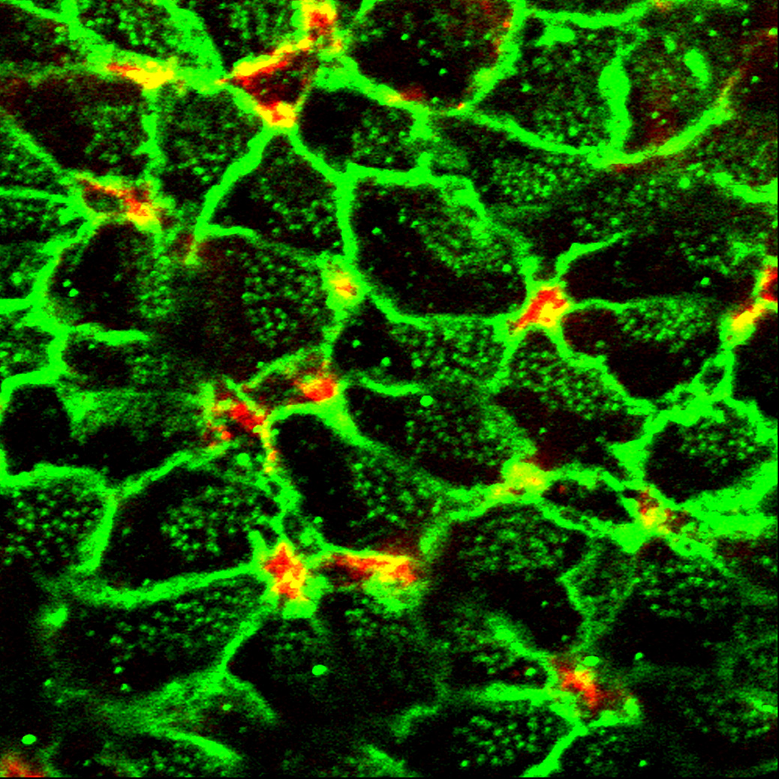
Figure 1. Neural stem cells in the SVZ
On the lateral wall of the lateral ventricle, neural stem cells (red) are surrounded by ependymal cells (green), resulting in a pinwheel-like arrangement.
Sonic hedgehog (Shh) is a critical signaling molecule that governs diverse developmental processes as well as postnatal neurogenesis in the brain. The Gli family of transcription factors (Gli1, Gli2, Gli3) are activated or modified in response to Shh activity. In particular, Gli3 is processed into a repressor form (Gli3R) in the absence of Shh signal and acts as the major negative transducer of the pathway. We have investigated the role of Gli3 prior to the onset of Shh signaling in the regulation of neural stem/progenitors in the developing brain and in the postnatal neurogenic niche.
The role of Gli3 in cortical development: Although the role of Gli3 in embryonic patterning has been extensively studied, its role in cortical development, especially in the regulation of proliferation and cell fate specification of neural progenitors, is largely unknown. When we conditionally remove Gli3 after the embryonic patterning is complete in mice, resulting Gli3 conditional mutants have enlarged lateral ventricles of the forebrain and thinner cortex compared with controls. Our results from birth-dating experiments showed that upper-layer cortical neurons were not produced properly and there was a prolonged production of deeper layer cortical neurons in Gli3 conditional mutants. Further investigation with in utero electroporation experiments showed that the Gli3, specifically Gli3R, is critical for specifying the fate of cortical neurons that are generated following a stereotypical temporal order. Moreover, we found that Gli3–mutant cells became post-mitotic prematurely, leading to a depletion of proliferating neural progenitors by late gestational stages. Our findings indicate that Gli3 is required for maintaining the cortical progenitors in active cell cycle, suggesting that cells may acquire differentiated status as they turn off Gli3 expression during neurogenesis.
The role of Gli3 in postnatal neurogenesis: Around birth, embryonic neural stem cells (also known as radial glia cells) undergo a transition to form the postnatal neurognic niche in the SVZ of the lateral ventricle. Neural stem cells in the SVZ are in close contact with other types of cells, including ependymal cells. While the niche cells are known to provide environmental cues to regulate adult neurogenesis, little is known about how the neurogenic niche is initially established and maintained. Therefore, we investigated the signaling mechanism that underpins the production of distinct cell types in the neurogenic niche. We discovered that Gli3 repressor (Gli3R), a negative regulator of Shh pathway, is critical for establishment and maintenance of the postnatal neurogenic niche in the SVZ in mouse forebrain. We first found that radial glia cells require Gli3R during development to correctly specify postnatal ependymal cells and neural stem cells. Using either genetic mutants or electroporation to remove Gli3 function in vivo, we then showed that proper neurogenesis depends on the continuous presence of Gli3R in ependymal cells—as opposed to neural stem cells. We found that Gli3R maintains an appropriate level of Numb, a negative regulator of the well-characterized juxtacrine signal Notch pathway, in ependymal cells to influence neurogenic activity of neighboring neural stem cells. Our findings highlight the importance of restricted expression of negative regulators of Shh and Notch pathways (Gli3R and Numb, respectively) in ependymal cells for proper neurogenesis and provide a better understanding of the interplay between neural stem cells and their environment.
Gene expression profiling of adult neural stem cells and their niche
Click image to enlarge.
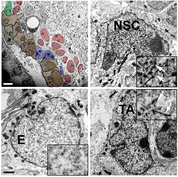
Figure 2. Shh-responding cells in the SVZ
Only neural stem cells (NSCs) and transit-amplifying (TA) cells, but not ependymal (E) cells, respond to Shh signaling and turn on Gli1 expression in the SVZ.
In order to understand the nature of the niche signals that regulate the behavior of neural stem cells in the SVZ of the lateral ventricle, we isolated highly purified neural stem cells and putative niche cells from the SVZ, including ependymal cells, astrocytes, and transit-amplifying cells, using flow cytometry, and from the choroid plexus by microdissection. We first validated the identity and purity of sorted cells by immunocytochemisty and confirmed the behavior of neural stem cells in vitro, i.e., their self-renewal and multi-potency. Next, we obtained the secretory-molecule expression profiling of each NSC niche cell type using the Signal Sequence Trap (SST) method. The resulting list contained genes that were previously implicated in the SVZ neurogenesis (cystatin C, ApoE) as well as newly discovered genes with unknown roles in the neurogenesis. We confirmed the expression of identified genes both in vitro (freshly sorted cells) and in vivo (tissue sections). We are currently assessing the physiological function of selected genes on neurogenesis in vitro. We are also establishing molecular and genetic networks among the secretory molecule expression profile (SMEP) by using bioinformatic tools such as Cytoscape and Ingenuity Pathway Analysis.
Sonic hedgehog signaling in dopamine neuron development
The ventral midbrain (vMb) is organized into distinct anatomical domains and contains cohorts of functionally distinct subtypes of midbrain dopamine (mDA) neurons. The diverse nature of mDA neurons in molecular identities, spatial and anatomical distribution, and neural circuitry governs the functional complexity of these neurons. We tested the hypothesis that genetic history and timing of gene expression within mDA neuron progenitors impart spatial diversity. Using genetic inducible fate mapping to mark the Shh and Gli1 lineages at varying embryonic stages, we performed a quantitative and qualitative comparison of the two lineages' contribution to the mDA neuron domains. Dynamic changes in Shh and Gli1 expression in the vMb primordia delineated their spatial contribution to the embryonic day 12.5 vMb; both lineages first contributed to the medial domain, but subsequently the Gli1 lineage exclusively contributed to the lateral vMb while the Shh lineage expanded more broadly across the vMb. The contribution of both lineages to the differentiated mDA neuron domain was initially biased anteriorly and became more uniform across the anterior/posterior vMb throughout development. Our findings demonstrate that the early Shh and Gli1 lineages specify mDA neurons of the substantia nigra pars compacta while the late Shh and Gli1 lineages maintain their progenitor state longer in the posterior vMb to extend the production of mDA neurons in the ventral tegmental area. Together, our study demonstrates that the timing of gene expression, along with the genetic lineage (Shh or Gli1) within the neural progenitors, segregates mDA neurons into distinct spatial domains.
Neural circuits formed by adult-born neurons
Neural circuit formation in aging brain: The hippocampus is another region of the brain that produces new neurons in adult animals. Neural stem cells reside in the subgranular zone of the dentate gyrus of the hippocampus and produce new granule neurons that project their axons to the CA3 region. In collaboration with Hwai-Jong Cheng, we are investigating how newborn neurons are incorporated into existing neural circuits of the hippocampus. Moreover, aging is known to slow down the rate of neurogenesis, but it is not known whether aging has any effects on integration of new neurons into the circuits. Using Gli1-CreER and Tau-mGFP mice to genetically label the neural stem cells in the hippocampus, we are visualizing the axonal projections through the detection of membrane-bound GFP reporter expression. We compared the labeled new neurons at various ages and analyzed the differentiation and maturation processes using serial immuno-elecronmicroscopy and three-dimensional reconstruction. Our preliminary results indicate that developmental integration of mossy fiber synaptic structures is compromised in the aged hippocampus.
Generation of novel circuitry reporter mice: Temporal and spatial control of marking cells of specific genetic lineages provides valuable insights into gene function during development and maturation processes. We developed Gli1-FlpePR-CreER(T2), a dual Genetic Inducible Fate Mapping (GIFM) system that allows us to control recombination events at two distinct time points with Tamoxifen and RU486. In addition, we are currently characterizing two universal reporter mouse lines that conditionally express fluorescent protein-tagged wheat germ agglutinin (tWGA:FP), which can be transferred across synapses during neural activities. Combinatorial use of our dual GIFM with the novel trans-synaptic reporter system enables us to trace the neural circuits that are formed between adult-born granule neurons from Shh-responding neural stem cells and pyramidal neurons of CA3 and subsequently of CA1.
Additional Funding
- DOD Grant (2009-2011)
Publications
- Wang H, Ge G, Uchida Y, Luu B, Ahn S. Gli3 is required for maintenance and fate specification of cortical progenitors. J Neurosci 2011;31:6440-6448.
- Hayes L, Zhang Z, Albert P, Zervas M, Ahn S. The timing of Sonic hedgehog and Gli1 expression segregates midbrain dopamine neurons. J Comp Neurol 2011;519:3001-3018.
- Chau M, Forcinito P, Andrade AC, Hegde A, Ahn S, Lui JC, Baron J, Nilsson O. Organization of the Indian hedgehog–parathyroid hormone-related protein system in the postnatal growth plate. J Mol Endocrinol 2011;47:99-107.
- Carney RSE, Mangin J-M, Hayes L, Mansfield K, Sousa V, Fishell G, Machold R, Ahn S, Gallo V, Corbin J. Sonic hedgehog expressing and responding cells generate neuronal diversity in the medial amygdala. Neural Dev 2010;5:14.
- Boudin CM, Gritli-Linde A, Ahn S, Harfe BD. Shh pathway activation is present and required within the vertebrate limb bud apical ectodermal ridge for normal autopod patterning. Proc Natl Acad Sci USA 107;5489-5494.
Collaborators
- Regina Armstrong, PhD, Uniformed Services University of the Health Service, Bethesda, MD
- Yosuke Mukoyama, PhD, Genetics and Developmental Biology Center, NHLBI, Bethesda, MD
- Mark Zervas, PhD, Brown University, Providence, RI
- Michael J. Holtzman, MD, Washington University, St. Louis, MO
- Hwai-Jong Cheng, MD, PhD, University of California, Davis, CA