Immune Activation and Viral Pathogenesis

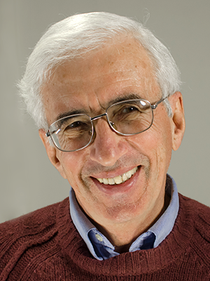
- Leonid Margolis, PhD, Head, Section on Intercellular Interactions
- Anush Arakelyan, PhD, Staff Scientist
- Christophe Vanpouille, PhD, Staff Scientist
- Rogers Nahui Palomino, BA, Visiting Fellow
- Wendy Fitzgerald, BS, Biologist
- Vincenzo Mercurio, MS, Predoctoral Visiting Fellow
The general goal of the Section for Intercellular Interactions is to understand the tissue-pathogenic mechanisms of human pathogens and the role of cytokines in such processes. We focused on the pathogenesis of human immunodeficiency virus 1 (HIV-1) and on its co-pathogens, in particular herpesviruses. We found that HIV-1 virions may carry the cytokine TGF beta to their cell target, probably facilitating HIV-1 infection. Also, the cytokine spectrum in semen appears to be an important determinant of HIV-1 transmission in vivo. Soluble and extracellular vesicle (EV)–associated cytokines are linked to the development of cardiovascular diseases, in particular of atherosclerosis triggered by HIV-1 infection. We also launched a project on SARS CoV-2 pathogenesis in human lung tissue ex vivo.
Macrophage-derived HIV-1 carries TGF-beta.
HIV-1 virions released by productively infected cells, predominantly T cells and macrophages, contain not only virus-encoded proteins but also some cellular proteins. The functions of the majority of cell-encoded molecules incorporated into virions, unlike those of virus-encoded molecules, are mostly not known, even though they may play an important role in HIV infection.
We focused on HIV-1 virions, produced by macrophages, given that in vivo these cells are long-term producers of viruses and probably dominate in the late stages of HIV disease. We used the single-virion analysis (“flow virometry”) that was developed in our laboratory several years ago and found that some of the HIV-1 virions produced by macrophages, along with viral antigens, incorporate into the viral membrane a macrophage scavenger receptor CD36.
We found that some CD36+ virions isolated from plasma of HIV–infected individuals are associated with transforming growth factor beta (TGF-beta), one of the key cytokines regulating immune responses. TGF-beta was found only on CD36+ virions derived from macrophages, not on CD27+ virions derived from T cells. TGF-beta binds to CD36 via thrombospondin-1(TSP-1), the main activator of TGF-beta. We proved that TGF-beta bound to macrophage-derived HIV remains bioactive, as shown using reporter cells.
Thus, HIV-1 not only induces general immune responses to infection but already carries cytokines that may affect the immune response, specifically in target cells. Viral-associated cytokines, together with cytokines produced by infected tissues, may determine a significant aspect of HIV-1 infection, including transmission, in particular sexual transmission.
Our work for demonstrated the first time that HIV virions can be associated with cytokines, namely TGF-beta.
Cytokines in HIV-1 transmission
A stated above, viral-associated cytokines, together with cytokines produced by infected tissues, may determine HIV-1 infection, in particular its sexual transmission. To address this question, we compared the cytokine/chemokine profiles in the blood and semen of source partners to investigate whether the profiles are associated with HIV transmission in the men’s recent sexual partners, who either became infected with a phylogenetically linked HIV strain (where the putative source is referred to as a transmitter) or did not acquire HIV (where the putative source partner is referred to as a non-transmitter). Viral transmission was confirmed by phylogenetic linkage (HIV pol). Using the multivariate statistical technique of partial least square discriminant analysis, we compared cytokine profiles of transmitters with those of non-transmitters. We found that the cytokine profiles of transmitters and non-transmitters were statistically different in semen but not in blood, suggesting that blood cytokines were not a good predictor of HIV transmission in our cohort mostly of men who have sex with men (MSM). There was a trend of higher concentrations of pro-inflammatory cytokines (RANTES, IL-18, IL-6, and GRO-alpha) in the blood of transmitters than in that of non-transmitters, but the overall difference was not statically significant.
In contrast to blood, the cytokine profiles in the semen of transmitters and non-transmitters were different, and the difference was highly significant. The finding reflects the fact that semen is not just a vector for HIV, but a complex milieu that carries pro- and antiviral factors that may facilitate or inhibit HIV transmission. The unique cytokine profile in the semen of the transmitter group suggests that seminal cytokines are likely to be an important determinant of HIV transmission. The differences in seminal cytokine profiles associated with transmission or non-transmission of HIV from infected MSM to their partners was still evident after the cytokine concentrations were adjusted for semen HIV RNA and cytomegalovirus (CMV) DNA.
We found no cytokines in semen that were associated with a higher risk of transmission. Rather, we found an associated group of cytokines: IFN-gamma, IL-13, M-CSF, IL-17, and GM-CSF. All cytokines except IL-13 were found in higher concentrations in non-transmitters than in transmitters. IFN-gamma and IL-13, the two cytokines with the highest differences, belong to different functional helper T cell type groups: IFN-gamma is the archetypal cytokine of the helper T cells type 1 (Th1) cells and supports cytotoxic T-cell responses, while IL-13 (together with IL-4) is secreted by helper T cells type 2 (Th2) cells, which counter-regulate Th1 responses and activate humoral immunity. A Th1/Th2 imbalance was originally described as a critical step in the etiology of HIV infection. Th1–associated cytokines have protective effects against HIV infection, whereas a shift towards an augmented humoral Th2 response may be detrimental and lead to the progression of HIV infection to acquired immunodeficiency syndrome.
Our study provides the first correlations between seminal cytokines in the transmitting partner and HIV transmission. The seminal cytokine spectrum is a contributing determinant of sexual HIV transmission, thus providing new directions for the development of strategies aimed at preventing HIV transmission.
Cytokines in HIV-1–triggered atherosclerosis
Dysregulation of cytokines in HIV-1–uninfected individuals is associated with T cell migration into atherosclerotic plaques. We showed that cytokines are released in both soluble and extracellular vesicle (EV)–associated forms. We investigated the expression and clustering of soluble and EV–associated cytokines in patients with ST-elevation myocardial infarction (STEMI). We found that several clustered cytokines were expressed almost exclusively in STEMI patients and were released in a coordinated fashion. Identification of such cytokine clusters permits investigation of their distinct contributions to STEMI. The cytokine pattern in STEMI patients resembles that triggered by viruses.
Although the etiological factor for atherosclerosis is not known, one such case is established: long-term HIV infection. In collaboration with a Case Western University team, we investigated the mechanisms by which cytokines trigger T cell recruitment to, and activation within, plaques. We found elevated expression of CX3CL1 (also known as fractalkine) in atherosclerotic plaques. The cytokine is known for eliciting its adhesive and migratory functions by interacting with the chemokine receptor CX3CR1, which we found to be upregulated on CD8+ T cells. Thus, endothelial cell–derived CX3CL1 may direct the migration of CX3CR1–expressing CD8+ T cells to the activated endothelium. These findings, together the observation that another elevated cytokine, IL-15, a strong activator of CD8+ T cells, induced expression of cytolytic molecules by the migrated CD8+ T cells, provide a mechanism for the damage caused to endothelia in the aortas of SIV- or SHIV–infected rhesus macaques.
In confirmation that a similar mechanism of atherosclerosis operates in humans, we found increased number of CD8+ T cells in atherosclerotic vessels of HIV–uninfected individuals. CD8+ T cells that accumulate in human atherosclerotic plaques have an activated, resident phenotype consistent with in vivo IL-15 and CX3CL1 exposure. Together these observations provide a novel model for CD8+ T cell involvement in atherosclerosis: CX3CL1 and IL-15 operate in tandem within the vascular endothelium to promote infiltration by activated CX3CR1+ memory CD8+ T cells, leading to atherosclerosis progression. Such processes are further promoted by the common HIV-1 co-pathogen CMV. The new results thus constitute newly discovered mechanisms of T cell infiltration into atherosclerotic plaques linked to cytokine disbalance.
SARS CoV-2 tissue pathogenesis
SARS CoV-2 infects lung epithelia, triggering severe pathology. Investigation of the mechanisms of SARS CoV-2 tissue pathogenesis and development of antiviral strategies in particular require a deep understanding of SARS CoV-2 pathogenesis in the context of human tissue systems under laboratory-controlled conditions. We developed such a system. Human lung tissue is dissected into 2mm3 blocks and cultured at the air-liquid interface. The histology of the blocks revealed well-preserved structural elements, including alveoli with epithelial cells. Flow cytometry of cells from these blocks confirmed their viability and expression of the ACE-2 receptor. To prove that the lung explants can be infected with SARS CoV-2, we used SARS CoV-2 pseudoviruses expressing the SARS-CoV-2 S protein that mediates cell infection with the virus. This pseudovirus encodes eGFP, thus making the infected cells fluoresce and allowing us to monitor viral infection. We tested the SARS CoV-2 pseudoviruses for infectivity on cell lines expressing ACE-2 and on lung tissues, and they were proven to be infectious. Development of human lung-tissue ex vivo pseudoviruses now allows us to characterize the main target cells of SARS CoV-2 with high-parameter flow cytometry.
Additional Funding
- Office of AIDS Research (OAR), NIH, Intramural Award
Publications
- Mercurio V, Fitzgerald W, Molodtsov I, Margolis L. Persistent immune activation in HIV-1-infected ex vivo model tissues subjected to antiretroviral therapy: soluble and extracellular vesicle-associated cytokines. J Acquir Immune Defic Syndr 2020;84:45.
- Ñahui Palomino RA, Vanpouille C, Laghi L, Parolin C, Melikov K, Backlund P, Vitali B, Margolis L. Extracellular vesicles from symbiotic vaginal lactobacilli inhibit HIV-1 infection of human tissues. Nat Commun 2019;10:5656.
- Lebedeva A, Fitzgerald W, Molodtsov I, Shpektor A, Vasilieva E, Leonid Margolis. Differential clusterization of soluble and extracellular vesicle-associated cytokines in myocardial infarction. Sci Rep 2020;10:21114.
- Bhatti G, Romero R, Rice GE, Fitzgerald W, Pacora P, Gomez-Lopez N, Kavdia M, Tarca AL, Margolis L. Compartmentalized profiling of amniotic fluid cytokines in women with preterm labor. PLoS One 2020;15:e0227881.
- Arakelyan A, Petersen JD, Blazkova J, Margolis L. Macrophage-derived HIV-1 carries bioactive TGF-beta. Sci Rep 2019;19100.
Collaborators
- Michael Bukrinsky, MD, PhD, George Washington University, Washington, DC
- Leonid Chernomordik, PhD, Section on Membrane Biology, NICHD, Bethesda, MD
- Sara Gianella Weibel, MD, University of California San Diego, La Jolla, CA
- Michael Freeman, PhD, Case Western University, Cleveland, OH
- Sergey Kochetkov, PhD, Engelhard Institute of Molecular Biology, Moscow, Russia
- Michael Lederman, MD, Case Western University, Cleveland, OH
- Roberto Romero-Galue, MD, DMedSci, Perinatology Research Branch, NICHD, Detroit, MI
- Yoel Sadovsky, MD, Magee-Womens Research Institute, University of Pittsburgh, Pittsburgh, PA
- Alexandr Shpektor, MD, Moscow Medical University, Moscow, Russia
- Elena Vasilieva, MD, Moscow Medical University, Moscow, Russia
- Beatrice Vitali, PhD, Università di Bologna, Bologna, Italy
Contact
For more information, email margolis@helix.nih.gov or visit http://irp.nih.gov/pi/leonid-margolis.


