Mechanisms Regulating GABAergic Cell Development

- Timothy J. Petros, PhD, Head, Unit on Cellular and Molecular Neurodevelopment
- Yajun Zhang, BM, Biologist
- Dongjin Lee, PhD, Postdoctoral Fellow
- Jianing Li, PhD, Postdoctoral Fellow
- Matthew Manion, PhD, Postdoctoral Fellow
- Chris Rhodes, PhD, Postdoctoral Fellow
- Dhanya Asokumar, BS, Postbaccalaureate Fellow
- Samra Beyene, MS, Postbaccalaureate Fellow
- Lielle Elisha, BS, Postbaccalaureate Fellow
- Anna Kim, Student Internship Program
The incredible diversity and heterogeneity of interneurons was observed over a century ago, with Ramon y Cajal hypothesizing in “Recollections of My Life” that “The functional superiority of the human brain is intimately linked up with the prodigious abundance and unaccustomed wealth of the so-called neurons with short axons.” Although interneurons constitute the minority (20%) of neurons in the brain, they are the primary source of inhibition and are critical components in the modulation and refinement of the flow of information throughout the nervous system. Abnormal development and function of interneurons has been linked to the pathobiology of numerous brain diseases, such as epilepsy, schizophrenia, and autism. Interneurons are an extremely heterogeneous cell population, with distinct morphologies, connectivities, neurochemical markers, and electrophysiological properties. With the advent of new technologies such as single-cell sequencing to dissect gene expression and connectivity patterns, the classification of interneurons into specific subtypes is ever evolving.
Interneurons such as GABAergic projection neurons are born in the ventral forebrain during embryogenesis and undergo a prolonged migratory period to populate nearly every brain region. However, our general understanding of the developmental mechanisms that generate such GABAergic cell diversity remains poorly understood. The goal of our lab is to dissect the genetic and molecular programs that underlie initial fate decisions during embryogenesis and to explore how the environment and genetic cascades interact to give rise to such a stunning diversity of GABAergic cell subtypes. We take a multifaceted approach, utilizing both in vitro and in vivo strategies to identify candidate mechanisms that regulate interneuron fate decisions. We strive to develop cutting-edge techniques that will overcome the many challenges faced when studying interneuron development. We believe that our pursuits will act as a springboard for future research and provide new insights into both normal development and various neurodevelopmental diseases.
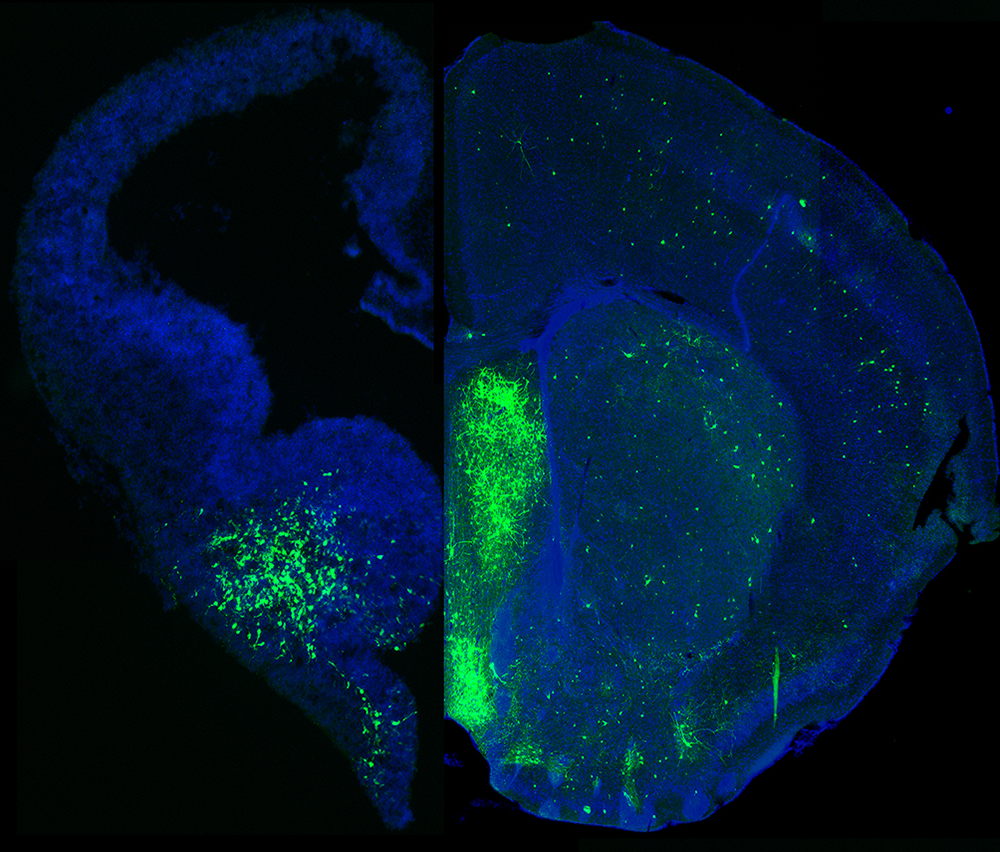
Figure 1. MGE–derived GABAergic cells populate many different brain regions.
Click image to view.
The image depicts a section of an embryonic brain (left) that has been electroplated to label cells derived from the medial ganglionic eminence (MGE), merged with a section of an adult brain (right), displaying the incredible spatial and morphological diversity of MGE–derived cells in the mature brain. Understanding how this heterogeneous population is generated from one embryonic brain structure is the focus of this laboratory.
Transcriptional heterogeneity of cycling VZ and SVZ cells throughout the embryonic mouse forebrain
The medial ganglionic eminence (MGE) gives rise to the majority of forebrain interneurons, most notably the somatostatin- and parvalbumin-expressing (SST+ and PV+) subtypes, and nNOS (neuronal nitric oxide synthetase)–expressing neurogliaform and ivy cells in the hippocampus. The MGE is a transient, dynamic structure, which arises around E10 and bulges into the lateral ventricle over the next several days before dissipating towards the end of embryogenesis. Given that initial fate decisions are generated within the MGE, there has been much focus on identifying a logic for interneuron generation from this region. Previous experiments characterized both a spatial and temporal gradient within the MGE, which regulates the initial fate decision to become either PV+ or SST+ interneurons. SST+ interneurons are preferentially born early in embryogenesis from the dorsoposterior MGE, whereas PV+ interneurons are born throughout embryogenesis with a bias of originating from the ventroanterior MGE. Earlier, I had discovered an additional mechanism regulating this fate decision: the mode of neurogenesis. Using in utero electroporations, I found that PV+ interneurons are preferentially born from basal progenitors (also known as intermediate progenitors), whereas SST+ interneurons arise more commonly from apical progenitors.
Recently, we built on this observation to characterize heterogeneous gene expression in neural progenitors throughout the embryonic forebrain. We performed a comprehensive single-cell RNA sequencing (scRNA-Seq) analysis of ventricular zone (VZ) and subventricular zone (SVZ) cells in four brain regions that give rise distinct cell types: the MGE, LGE (lateral ganglionic eminence), CGE (caudal ganglionic eminence), and cortex. This allows us to compare gene expression in neural progenitors both between distinct brain regions (MGE vs. CGE) and within specific subdomains of these regions (dorsal vs. ventral LGE). We verified many of these gene expression profiles via in situ hybridization and revealed previously unknown transcriptional heterogeneity in VZ cells throughout the forebrain. The findings were published earlier this year [Reference 2]. We are currently following up on several intriguing candidates to better understand their role in cell fate.
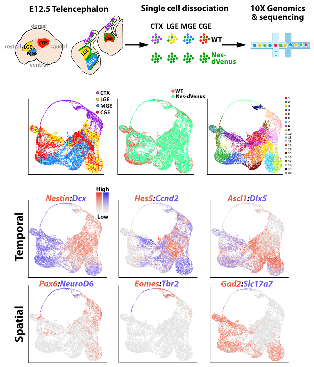
Figure 2. Single-cell sequencing in the embryonic mouse forebrain
Click image to view.
Top row. Experimental paradigm to harvest cells from four distinct brain regions (MGE, LGE, CGE, cortex) from wild-type and Nestin-dVenus embryonic mouse brains for single-cell RNA sequencing.
Middle row. UMAP (uniform manifold approximation and projection) plots of single cells categorized by brain region (left), mouse line (middle), or putative cell cluster (right).
Bottom rows. UMAP plots depicting gene expression profiles enriched in different stages of neural progenitors (top) or spatial domains (bottom).
Characterization of the epigenetic landscape during embryonic neurogenesis
In multicellular organisms, cells are genetically homogenous but structurally and functionally heterogeneous as a result of differential gene expression, which is often mitotically heritable. The mechanisms regulating such expression are ‘epigenetic,’ as they do not involve altering the DNA sequence itself; they include DNA methylation (DNAme), histone modifications, and higher-order chromatin structure. In particular, DNA and histone modifications often follow specific rules termed the ‘epigenetic code,’ similar to the genetic code. Collectively, DNAme and histone modification have been reported to regulate transcription and chromatin structure in many stem-cell and developmentally critical processes. Previous scRNA-Seq experiments on the ganglionic eminences (GEs) identified surprisingly few region-specific genes in cycling progenitors (immature cells that are still cycling and have not exited the cell cycle), despite the fact that these regions produce distinct GABAergic cell populations. Because there are dynamic changes in the chromatin landscape during development, a prevailing hypothesis is that epigenomic signatures may be a better predictor of cell fate during development, revealing both potential distal enhancers and/or genetic loci that may be ‘poised’ but not yet expressed. However, direct support for this hypothesis is lacking. The idea is particularly relevant, given that epigenetic changes are observed in many neurological and psychiatric diseases and that most single-nucleotide variants (SNVs) identified in diseases-specific genome-wide association studies (GWAS) map to non-coding regions, implying that epigenetic regulation of gene expression may underlie some disease etiologies.
We performed single-cell assay for transposase-accessible chromatin with sequencing (scATAC-Seq), in combination with Cut&Tag (to characterize histone modifications) and Hi-C/Capture-C (to analyze higher order chromatin interactions), in order to generate an ‘Epigenome Atlas’ of four different regions in the embryonic mouse brain. This comprehensive, unbiased analysis reveals striking differences in gene expression and chromatin organization between adjacent brain regions and revealed new candidates for region-specific promoter-enhancer interactions that may be critical for neurons' fate and maturation. We are currently following up on several intriguing avenues based on these observations. The work was published earlier this year [Reference 1], and the entire dataset is publicly available in a searchable format on the UCSC Genome Browser platform. Additionally, based on this study and the one above, we recently prepared a manuscript detailing our procedures to obtain single-cell and single-nuclei dissociations from embryonic and adult mouse brains for numerous downstream applications [Reference 3].
Figure 3. snATAC-Seq in distinct regions of the mouse embryonic forebrain

Click image to view.
A. Schematic of snATAC-Seq (single-nucleus analysis of transposase-accessible chromatin using sequencing) workflow and neurogenic cell types: apical progenitors (APs), basal progenitors (BPs) and neurons (Ns).
B. UMAP (uniform manifold approximation and projection) visualization of single nuclei clustered by brain region;
C. SLM (Smart Local Moving algorithm);
D. neurogenic cell type. PA (promoter accessibility) representing reads mapping within 2 kb upstream of TSSs (transcription start sites);
E. pseudotime;
F. Embryonic snATAC-Seq dot plot of differentially accessible peaks (DA peaks) for each cluster. Dot diameter indicates the percent of DA peaks from one cluster (column cluster labels) that are detectable in any other cluster (row cluster labels). Color intensity represents the total DA peak count per cluster. Hierarchical clustering was performed using correlation distance and average linkage.
Mechanisms regulating fate determination of CGE–derived interneurons
While significant progress has been made characterizing mechanisms regulating initial fate decisions of MGE–derived interneurons, our understanding of CGE–derived interneurons lags significantly. This is in part because we lack genetic tools to specifically target and manipulate CGE–derived cells. There is an expansion of CGE–derived interneuron subtypes in humans and primates compared with mice, so a better understanding of the developmental trajectory of these cells is warranted.
To this end, we are currently performing two types of experiments to better understand the developmental logic of CGE–derived cells. First, we are performing cell transplantation studies to determine whether specific CGE–derived interneuron subtypes are derived from distinct regions of the CGE. These types of transplantation experiments generated initial insights into the spatial logic of the MGE, but surprisingly similar types of experiments have not been performed in the CGE. By combining this spatial logic with the scRNA-Seq and scATAC studies detailed above, we hope to link early transcription and chromatin accessibility profiles in CGE progenitors with mature interneuron fates. Second, we are developing a Perturb-Seq approach using mESCs to identify activation (or repression) of genes that promote a CGE fate. We hope to make significant progress in both these approaches in the upcoming year.
Publications
- Rhodes CT, Thompson JJ, Mitra A, Asokumar D, Lee DR, Lee DJ, Zhang Y, Jason E, Dale RK, Rocha PP, Petros TJ. An epigenome atlas of neural progenitors within the embryonic mouse forebrain. Nat Commun 2022 13(1):4196.
- Lee DR, Rhodes CT, Mitra A, Zhang Y, Maric D, Dale RK, Petros TJ. Transcriptional heterogeneity of ventricular zone cells in the ganglionic eminences of the mouse forebrain. eLife 2022 https://doi.org/10.7554/eLife.71864.
- Lee DR, Zhang Y, Rhodes CT, Petros TJ. Generation of single cell and single nuclei suspensions from embryonic and adult mouse brains. STAR Protocols 2022 4(1):101944.
- Chakraborty S, Kopitchinski N, Zuo Z, Eraso A, Awasthi P, Chari P, Mitra A, Tobias IC, Moorthy SD, Dale RK, Mitchell JA, Petros TJ, Rocha PP. Enhancers that activate target genes across CTCF boundaries increase phenotypic robustness. Nat Genet 2022 in press.
Collaborators
- Susan Amara, PhD, Laboratory of Molecular and Cellular Neurobiology, NIMH, Bethesda, MD
- Ryan Dale, PhD, Bioinformatics Core, NICHD, Bethesda, MD
- Claire Le Pichon, PhD, Unit on the Development of Neurodegeneration, NICHD, Bethesda, MD
- Chris McBain, PhD, Section on Cellular and Synaptic Physiology, NICHD, Bethesda, MD
- Pedro Rocha, PhD, Unit on Genome Structure and Regulation, NICHD, Bethesda, MD
Contact
For more information, email tim.petros@nih.gov or visit https://www.nichd.nih.gov/research/atNICHD/Investigators/petros.