Quantitative Imaging and Tissue Sciences

- Peter J. Basser, PhD, Head, Section on Quantitative Imaging and Tissue Sciences
- Ferenc Horkay, PhD, Staff Scientist
- Alexandru Avram, PhD, Research Fellow (via NIBIB)
- Magdoom Kulam, PhD, Postdoctoral Visiting Fellow
- Matan Mussel, PhD, Postdoctoral Visiting Fellow
- Nathan Hu Williamson, PhD, Postdoctoral Intramural Research Training Award Fellow
- Velencia Witherspoon, PhD, Postdoctoral Intramural Research Training Award Fellow
- Adam Bernstein, PhD, Predoctoral Intramural Research Training Award Fellow
- Teddy Cai, BS, Predoctoral Intramural Research Training Award Fellow, NIH OxCam Program
- Dan Benjamini, PhD, Collaborating Scientist funded via the Henry Jackson Foundation and the Center for Neuroscience and Regenerative Medicine
- Michal Komlosh, PhD, Collaborating Scientist funded via the Henry Jackson Foundation and the Center for Neuroscience and Regenerative Medicine
- Kadharbatcha Saleem, PhD, Collaborating Scientist funded via the Henry Jackson Foundation and the Center for Neuroscience and Regenerative Medicine
- Alexandros Chremos, PhD, Contract Scientist
- Rea Ravin, PhD, Contract Scientist
In our tissue sciences research, we strive to understand fundamental relationships between function and structure in living tissues, using 'engineered' tissue constructs and tissue analogs. Specifically, we are interested in how microstructure, hierarchical organization, composition, and material properties of tissues affect their biological function or dysfunction. We investigate biological and physical model systems at various length and time scales, performing physical measurements in tandem with developing physical/mathematical models to explain their functional properties and behavior. Experimentally, we use water to probe both equilibrium behavior and dynamic interactions among tissue constituents from nanometers to centimeters and from microseconds to lifetimes. To determine the equilibrium osmo-mechanical properties of well-defined model systems, we vary water content or ionic composition systematically. To probe tissue structure and dynamics, we also employ atomic force microscopy (AFM), small-angle X-ray scattering (SAXS), small-angle neutron scattering (SANS), static light scattering (SLS), dynamic light scattering (DLS), and one- and two-dimensional nuclear magnetic resonance (NMR) relaxometry and diffusometry. A goal of our basic tissue sciences research is to develop understanding and tools that can be translated from bench-based quantitative methodologies to the bedside.
Our tissue sciences activities dovetail with our basic and applied research in quantitative imaging, which is intended to generate measurements and maps of intrinsic physical quantities, including diffusivities, relaxivities, or exchange rates, rather than the qualitative stains and images conventionally used in radiology. At a basic level, the work is directed toward making invisible structures and processes visible. Our quantitative imaging group uses knowledge of physics, engineering, applied mathematics, imaging, and computer sciences, as well as insights gleaned from our tissue-sciences research to discover and develop novel imaging biomarkers that can detect changes in tissue composition, microstructure, or microdynamics more sensitively and specifically. The ultimate translational goal is to be able assess normal and abnormal development, diagnose childhood diseases and disorders, and characterize degeneration and trauma. Primarily, we use MRI as our imaging modality of choice because it is so well-suited to many applications critical to NICHD's mission; it is noninvasive, nonionizing, requires (in most cases) no exogenous contrast agents or dyes, and is generally deemed safe and effective for use with fetuses and children in both clinical and research settings.
A technical objective has been to transform clinical MRI scanners into scientific instruments capable of producing reproducible, highly accurate, and precise imaging data with which to measure and map useful imaging quantities for various clinical applications, including single scans, longitudinal and multi-site studies, personalized medicine, and genotype/phenotype correlation studies, as well as for populating imaging databases with high-quality normative data. We also develop our various MRI tools and methodologies to advance the field of neuroscience, providing means to explore brain structure and function.
Click image to view.
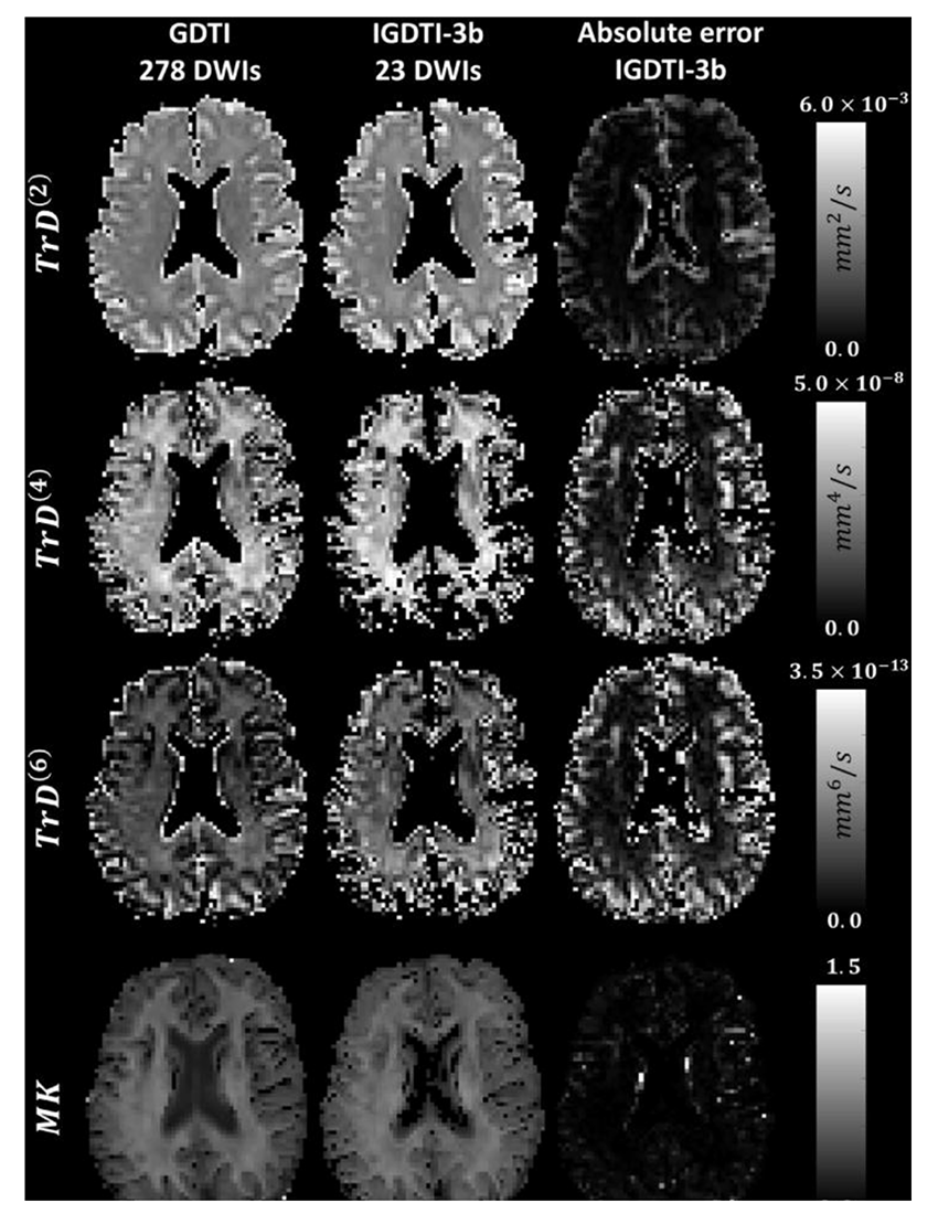
Figure 1. New isotropically weighted diffusion MRI method shows novel anatomical features in living human brain.
Comparison of orientationally averaged Trace of diffusion tensors, TrD(n), for tensors of orders 2, 4, and 6, and mean t-kurtosis, W¯ measured in vivo with Generalized Diffusion Tensor Imaging (GDTI) and Isotropic Generalized Diffusion Tensor Imaging (IGDTI) with 3 b-values. Figure shows the ability of IGDTI to quantify rotation-invariant higher-order tensor (HOT)–derived diffusion parameters in brain tissue within a clinically feasible scan duration.
In vivo MRI histology
The most mature in vivo MRI technology that we invented, developed, and clinically translated is Diffusion Tensor MRI (DTI), by which we measure D, a diffusion tensor of water, voxel-by-voxel within an imaging volume. Information derived from this quantity includes white-matter fiber-tract orientation, the mean-squared distance that water molecules diffuse in each direction, the orientationally averaged mean diffusivity, and other intrinsic scalar (invariant) quantities. These imaging parameters have been used by radiologists and neuroscientists as non-invasive quantitative histological 'stains'. Remarkably, the MRI images are obtained by probing endogenous tissue water in vivo without requiring exogenous contrast agents or dyes. The bulk or orientationally averaged apparent diffusion coefficient (mean ADC) is the most successful and widely used DTI parameter to identify ischemic regions in the brain during acute stroke and to follow cancer patients' response to therapy. Our measures of diffusion anisotropy (e.g., fractional anisotropy or FA) are used universally to follow changes in normally and abnormally developing white matter, including dysmyelination and demyelination and many other applications of brain white matter visualization. Our group also pioneered the use of fiber direction–encoded color (DEC) maps to display the orientation of the main association, projection, and commissural white matter pathways in the brain. To assess anatomical connectivity among various cortical and deep-brain gray-matter areas, we also proposed and developed DTI "Streamline" Tractography. Collectively, these advances helped inspire several large federally funded research initiatives, such as the NIH Human Connectome Project (HCP)
More recently, we invented and developed a family of advanced in vivo diffusion MRI methods to measure fine-scale microstructural features of axons and fascicles, which otherwise could only be measured using laborious ex vivo histological methods. We have been developing efficient means for performing "k and q-space MRI" in the living human brain, such as "Mean Apparent Propagator" (MAP) MRI. This approach detects subtle microstructural and architectural features in both gray and white matter at micron-scale resolution, several orders of magnitude smaller than the typical MRI voxel. MAP-MRI also subsumes DTI, as well as providing a bevy of new in vivo quantitative 'stains' to measure and map. We also developed a family of diffusion MRI methods to 'drill down into the voxel' and measure features such as average axon diameter (AAD) and axon-diameter distribution (ADD) within and along large white-matter pathways, dubbing them CHARMED and AxCaliber MRI, respectively. After careful validation studies, we reported the first in vivo measurement of ADDs within the rodent corpus callosum. The ADD is functionally important, given that axon diameter is a determinant of axon or nerve conduction velocity and therefore the rate at which information flows along white matter pathways, helping to determine the delays or latencies between and among different brain areas. We then developed a companion mathematical theory to explain the observed ADDs in different fascicles, suggesting that they represent a trade-off between maximizing information flow and minimizing metabolic demands. We also developed novel multiple pulsed-field gradient (mPFG) methods and demonstrated their feasibility for use in vivo on conventional clinical MRI scanners as a further means to extract quantitative features to measure and map in the central nervous system (CNS). The methods can also provide an independent measurement of the AAD and other features of cell size and shape.
Although gray matter appears featureless in DTI brain maps, its microstructure and architecture are rich and varied throughout the brain, not only along the brain's cortical surface, but also within and among its various cortical layers and within deep gray-matter regions. To target this tissue, we have been developing several noninvasive, in vivo methods to measure unique features of cortical gray matter microstructure and architecture that are currently invisible in conventional MRI. One of our long-term goals is to 'parcellate' or segment the cerebral cortex in vivo into its approximately 500 distinct cyto-architechtonic areas. To this end, we are developing advanced MRI sequences to probe correlations among microscopic displacements of water molecules in the neuropil as well as sophisticated mathematical models to infer distinguishing microstructural and morphological features of gray matter. We pioneered several promising two-dimensional MRI relaxometry, exchange, and diffusometry-based methods to study water mobility and exchange in gray and white matter. We believe these will help identify changes in normal and abnormal development, as well as inflammation and trauma.
Quantitative fetal and pediatric MRI
MRI is considered safer than X-ray–based methods, such as computed tomography (CT), for scanning fetuses, infants, and children. However, clinical MRI still lacks the quantitative character of CT. Clinical MRI relies upon the acquisition of "weighted images," whose contrast is affected by many factors, some intrinsic to the tissue and some dependent on the details of the experiment and experimental design. It is also prone to more acquisition and imaging artifacts. The diagnostic utility of conventional MRI for many neurological disorders is unquestionable. However, the scope of conventional MRI applications is limited to revealing either gross morphological features or focal abnormalities, which result in regional differences in signal intensities within a given tissue. Clinical MRI also often lacks biological specificity. Although quantification per se does not ensure improved specificity, it is nonetheless necessary for developing robust and reliable imaging "biomarkers." In particular, MRI assessment of normal brain development and developmental disorders has benefited greatly from the introduction of "quantitative" clinical MRI techniques, with which one obtains maps of meaningful intrinsic physical quantities or chemical variables, which can be measured in physical units and compared among different tissue regions, in individual subjects, and within longitudinal and cross-sectional studies. Quantitative MRI methods, such as DTI, also increase sensitivity, providing a basis for monitoring subtle changes that occur, e.g., during the progression or remission of disease, by comparing measurements in a single subject with normative values obtained from a healthy population. Quantitative MRI methods should continue to advance "precision imaging" studies, in which MRI phenotypic and genotypic data can be meaningfully melded and used for improved diagnosis and prognosis assessments.
To date, we have developed algorithms that generate a continuous, smooth approximation to the discrete, noisy, measured DTI field data so as to reduce noise and allow us to follow fiber tracts more reliably. We proposed a novel Gaussian distribution for the tensor-valued random variables that we use in designing optimal DTI experiments and interpreting their results. In tandem, we developed nonparametric empirical (e.g., Bootstrap) methods to determine the statistical distribution of DTI–derived quantities in order to study, for example, the inherent variability and reliability of computed white-matter fiber-tract trajectories. Such parametric and nonparametric statistical methods enable us to apply powerful hypothesis tests to assess the statistical significance of findings in a wide range of important biological and clinical applications that are currently being tested using ad hoc statistical methods. We are also developing novel methods to register or warp different brain volumes and to generate group-average data or atlases from various subject populations based on the Kullback-Leibler divergence. However, much work remains to be done in order to address and remedy MRI artifacts so as to permit one to draw statistically significant inferences from clinical DTI data, obtained in longitudinal and multi-center studies, and particularly in single-subject studies.
We carry out key clinical studies that utilize novel quantitative MRI acquisition and analysis methods and whose aim is to improve accuracy and reproducibility in diagnosis and to detect and follow normal and abnormal development. One example is the NIH Study of Normal Brain Development, jointly sponsored by the NICHD, NIMH, NINDS, and NIDA. This multi-center consortium, initiated in 1998, was intended to advance our understanding of normal brain development in typical healthy children and adolescents. The Brain Development Cooperative Group, created by this funding mechanism, is still active, publishing numerous papers each year, primarily by mining these rich high-quality data, many of which we processed, vetted, and uploaded. We served as the DTI Data-Processing Center (DPC) in this interdisciplinary project. The processed DTI data collected from this project were uploaded into a database accessible to interested investigators, and made publicly available through the National Database for Autism Research (NDAR). In collaboration with Carlo Pierpaoli, we continue to support and help update and disseminate the processing and analysis software that grew out of this effort, called "TORTOISE," which can be downloaded from http://www.tortoisedti.org.
Our involvement in traumatic brain injury (TBI) research, particularly in detecting mild TBI (mTBI), has continued to expand and in partnership with various Department of Defense (DoD) affiliates. TBI is an acute problem in the pediatric population, but it also affects and afflicts young men and women in the military. Diffusion MRI provides essential information to aid in the assessment of TBI. Because of the subtlety of changes seen in TBI, quantitative imaging protocols are essential, as are the development of advanced imaging methods. To this end, we developed a DTI data–processing pipeline in order to improve the accuracy and reproducibility of MAP–MRI findings, and, in collaboration with scientists at the DoD Center for Neuroscience and Regenerative Medicine (CNRM), performed the first normative MAP–MRI studies, as well as applied this new and powerful method to individuals with mild and moderate TBI. To enable this application, we built on our existing state-of-the-art TORTOISE pipeline to include and incorporate MAP–MRI data. We are continuing to develop and use novel MRI phantoms, such as our polyvinyl pyrrolidone (PVP) polymer and Glass Capillary Array (GCA) MRI phantoms that we developed and are disseminating to improve the performance of advanced diffusion MRI methods.
In collaborative research fostered by our dynamic Affinity Group in the area of fetal MRI, an in collaboration with Roberto Romero, we are exploring ways to "freeze" the motion of fetal brains as the fetus moves throughout the scan. We are also developing approaches to characterize microstructure and function, initially in the fetal brain, but eventually within the entire placenta.
Biopolymer physics: water-ion-biopolymer interactions
Water-ion-biopolymer interactions play a key role in the physical-chemical interactions among tissue components. Our primary objective is to understand the forces that govern these interactions, ranging from the movement of water and ions across channels, to the self-assembly of proteins into nanofibers in neuro-degenerative processes, such as Creutzfeldt-Jakob disease, Alzheimer’s disease, and chronic traumatic encephalopathy (CTE). Little is understood about the physical underpinnings of these interactions, which underlie a myriad of biological processes and behaviors.
To determine the effect of ions on the structure and dynamics of biopolymers, we developed a multiscale experimental framework by combining macroscopic techniques (osmotic swelling pressure measurements, mechanical measurements) with scattering methods (e.g., SANS and SAXS), which probe the structure and interactions over a broad range of length and time scales. Macroscopic swelling pressure measurements provide information on the overall thermodynamic response of the system, while SANS and SAXS allow us to investigate biopolymers at the molecular and supramolecular length scales and to quantify the effect of changes in the environment (e.g., ion concentration, ion valance, pH, temperature) on the structure and interactions among biopolymers, water, and ions. Studies made on well-defined model systems that mimic certain essential features of tissue provide important insights that cannot be obtained from experimental studies made on biological systems. Mathematical models based on well-established polymer physics concepts makes it possible to design and analyze fundamental experiments, which quantify and explain tissue behavior. We are now combining molecular dynamics simulations with cutting-edge experimental studies to gain insight into the underlying molecular mechanism that governs key aspects of tissue structure-function relationships.
Better understanding of the structure and interactions among tissue components is also necessary to design and develop tissue models and novel tissue phantoms that mimic tissue behavior. Biomimetic phantoms with well-controlled physical properties (osmotic, mechanical, relaxation, etc.) are critically important in quantitative MRI to validate measurements and advanced MRI applications from bench to bedside (in vivo MRI histology). We produced novel diffusion MRI phantoms, which we use to calibrate MRI scanners, specifically to assure the quality of the imaging data and to assess scanner performance on an on-going basis. Our U.S. Patent for a "Phantom for diffusion MRI imaging" is enabling quantitative diffusion MRI studies to be performed at many different sites. The polymer consisting of the phantom has ideal properties for this demanding application. Colleagues at NIST in Boulder, Colorado, have incorporated our PVP polymer into NIST's own diffusion MRI standard. The technology is also being promulgated commercially e.g., by http://hpd-online.com/diffusion-phantom.php. We developed a variety of NMR and MRI phantoms that possess various salient features of cell or tissue systems, providing 'ground truth' to test the validity of our models and experimental designs.
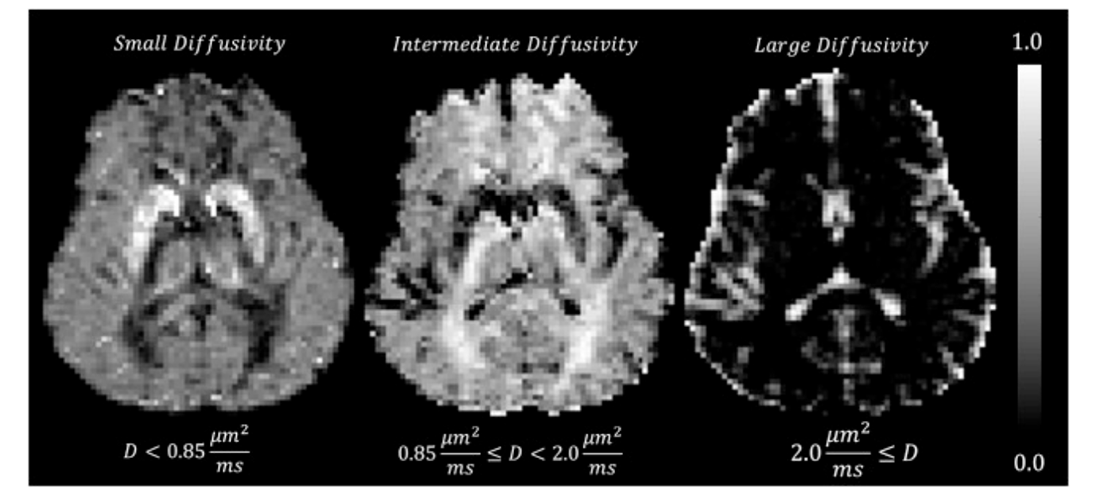
Click image to view.
Figure 2. Spectra of mean water diffusivities within living human brain
Low, intermediate, and large diffusivity signal components in normalized mean diffusion distributions (MDDs) measured in live human brain. Subcortical gray matter, in particular the putamen, the globus pallidus, the caudate nucleus, and, to a lesser extent the thalamus, contain mostly low diffusivity components ≤ 0.85 μm2/ms. Brain regions containing primary white matter (WM) pathways are dominated by intermediate diffusivity components in the range of 0.85−2.0 μm2/ms. Peaks in grey matter (GM) contain significant signal fractions of small and intermediate mean diffusivities. As expected, the largest diffusivity values are observed mainly in regions of the CSF (cerebrospinal fluid). This approach allows us to cleanly cluster different tissue types and assess the spectrum of mean water diffusivities within them.
Measuring and mapping functional properties of extracellular matrix
Extracellular matrix (ECM) is present in every tissue and performs a key role in determining normal and abnormal tissue and organ function. We study interactions among the main ECM components using cartilage as a model system. In cartilage ECM, the most abundant macromolecule is collagen (type II), which is organized into fiber bundles that form a meshwork. The major proteoglycan (PG) is the bottlebrush-shaped aggrecan. Intracellularly synthesized aggrecan molecules are secreted into the ECM, where they aggregate to form a secondary bottlebrush with hyaluronic acid (HA), which is stabilized by a link protein. The biomechanical behavior of cartilage and other ECMs reflects their molecular composition and microstructure, which change during development, disease, degeneration, and aging. To determine tissue structure/function relationships, we measure various physical/chemical properties of ECM tissues and tissue analogs at different length- and time-scales, using a variety of complementary static and dynamic experimental techniques, e.g., osmometry, SANS, SAXS, neutron spin-echo (NSE), SLS, DLS, AFM, and fluorescence correlation spectroscopy (FCS). Understanding the physical and chemical mechanisms affecting cartilage swelling (hydration) is essential to predicting cartilage load-bearing properties, which are mainly governed by osmotic and electrostatic forces. To quantify the effect of hydration on cartilage properties, we previously developed a tissue micro-osmometer to perform precise measurements in a rapid manner. The instrument is capable of determining very small changes in the amount of water absorbed by small tissue samples (less than 1 microgram) as a function of the equilibrium water activity (vapor pressure). We make osmotic pressure measurements to determine how the individual components of cartilage ECM (e.g., aggrecan, HA, and collagen) contribute to the total load-bearing capacity of the tissue. We demonstrated that aggrecan–HA aggregates self-assemble into microgels, contributing to improved dimensional stability and the tissue's lubricating ability. We also found that aggrecan is highly insensitive to changes in the ionic environment, particularly to the concentration of calcium ions, which is critically important to maintaining the tissue's mechanical integrity and to allowing aggrecan to serve as a calcium ion reservoir in cartilage and bone.
We developed a new biomimetic model of cartilage ECM, consisting of polyacrylic acid (PAA) microgel particles dispersed and embedded within a polyvinyl alcohol (PVA) gel matrix. In this composite system, PAA mimics the behavior of proteoglycan (i.e., HA–aggrecan complexes), while PVA mimics the behavior of the collagen network. The PVA/PAA biomimetic model system reproduces not only the shape of the cartilage swelling pressure curves, but also the numerical values reported for healthy and osteoarthritic human cartilage samples. Systematic studies made on model composite hydrogels is expected to provide invaluable insights into the effects of various factors (matrix stiffness, swelling pressure, fixed-charge density, etc.) on the macroscopic mechanical/swelling properties, and ultimately the origin of the load-bearing and lubricating ability of cartilage.
We are also using osmotic pressure measurements to determine the contributions of individual components of ECM to the total tissue swelling pressure. In collaboration with Emilios Dimitriadis, we developed a method for mapping the local elastic and osmotic properties of cartilage, using AFM together with the tissue micro-osmometer. Many of the impediments that previously hindered the use of AFM to probe inhomogeneous samples, particularly biological tissues, were addressed by this approach that utilizes the precise scanning capabilities of a commercial AFM to generate large volumes of compliance data, from which the relevant tissue elastic properties can be extracted. In conjunction with results obtained from high-resolution scattering measurements and biochemical analysis, the technique allows us to map the spatial variations in the osmotic modulus within tissue specimens and thus the compressive resistance of the tissue to external load.
We are attempting to translate this critical tissue-science understanding of the structure-function relationships of ECM components to develop and design novel non-invasive MR imaging methods, with the aim of inferring ECM composition, patency, and functional properties in vivo. Our goal is to use MRI for early diagnosis of diseases of cartilage and other tissue and organs, as well as to provide a means for following normal and abnormal development of the ECM, which entails making 'invisible' components of ECM, (e.g., collagen and PGs) 'visible' and then using our understanding of biopolymer interactions to predict functional properties of the tissue, such as its load-bearing ability. One major obstacle is that water molecules bound to immobile species (e.g., collagen) are largely invisible to conventional MRI approaches. However, magnetization exchange (MEX) MRI (as well as other methods) make it possible to detect the bound protons indirectly by transferring their magnetization to the free water surrounding them. It also enables us to estimate the collagen content in tissue. In a pilot study with collaborators Uzi Eliav and Ed Mertz, we applied the new MEX MRI method to determine the concentration and distribution of the main macromolecular constituents in bovine femoral-head cartilage samples. The results obtained by the MEX MRI method are qualitatively consistent with those obtained by histological techniques, such as high-definition infrared (HDIRI) spectroscopic imaging. The work was originally supported by a DIR Director's Award that we received with our collaborators Sergey Leikin and Edward Mertz, and are continuing to pursue collaboratively. Our novel approach has the potential to map tissue structure and functional properties in vivo and noninvasively, so while it is "high-risk" it has the potential for a "high-reward."
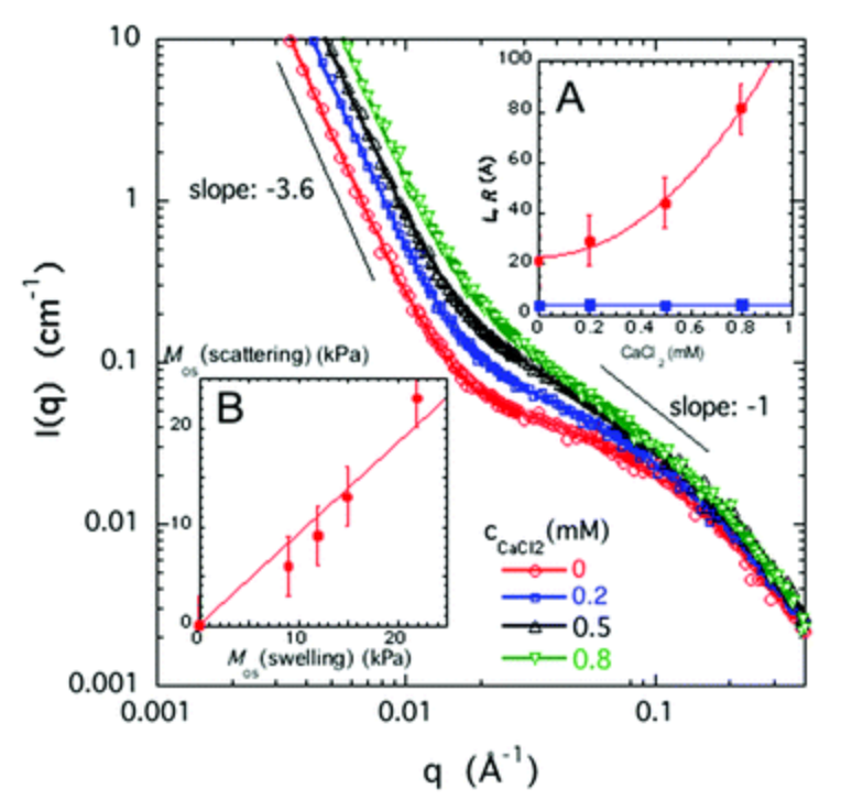
Click image to view.
Figure 3. Remarkable correspondence between load bearing ability of polyelectrolyte gels measure by Small Angle Neutron Scattering and Osmotic Stress techniques
Small-angle neutron scattering profiles of sodium polyacrylate (NaPA) gels (φ = 0.04) swollen in 40 mM NaCl containing increasing amounts of CaCl2 as indicated in the figure. Inset (A): Variation of the correlation length L (red dots) and the cross-sectional radius, R (blue squares) as a function of the CaCl2 concentration. SANS provides a powerful high-resolution method to probe molecular structure over a large range of length scales. Inset (B): Osmotic modulus, Mos(scattering) vs. osmotic modulus Mos(swelling) for NaPA gels. Notice the excellent agreement between the swelling and scattering measurements.
Patents
- Benjamini D, Basser PJ. Multi-dimensional spectroscopic NMR and MRI using marginal distributions. USPTO Patent No. WO2018031942-A1 2018;N/A.
- Horkay F, Pierpaoli C, Basser PJ. Phantom for diffusion MRI imaging. USPTO Patent No. 10,078,124 2018;N/A.
Additional Funding
- “Characterizing brain microstructure in patients with mTBI using Mean Apparent Propagator (MAP) MRI.” HJF Award Number: 308049-8.01-60855, (CNRM-89-3895), which is under the joint auspices of the NIH, DoD, CNRM, and USUHS.
- “Development of Bench and Pre-Clinical MRI Methods to Assess Glymphatic Clearance in the Living Brain.” 308811-4.01-60855, (CNRM-89-9237), which is under the joint auspices of the NIH, DoD, CNRM, and USUHS.
- “In vivo Brain Network Latency Mapping.” NIH BRAIN Initiative grant 1-R24-MH-109068-01
- “Connectome 2.0: Developing the next generation human MRI scanner for bridging studies of the micro-, meso- and macro-connectome”, NIH BRAIN Initiative-funded 1U01EB026996-01
- “MRI methods aimed at the detection of pathologies involving myelin, collagen and amyloid plaques” Bi-National Science Foundation (BSF) grant; 2013-2018, #2013253
- “Neuroradiology/Neuropathology Correlation/Integration Core”, 309698-4.01-65310, (CNRM-89-9921)
- “Localization of Epileptogenic Foci using Diffusion Weighted MRI”, Bench-to-Bedside (BtB) Award, NIH Director’s Challenge Innovation Grant, NIH IRP
Publications
- Horkay F, Basser PJ, Hecht AM, Geissler E. Microstructure and dynamic properties of aggrecan assemblies. MRS Adv 2018;3(28):1589-1595.
- Avram A, Sarlls J, Basser PJ. Measuring non-parametric distributions of intravoxel mean diffusivities using a clinical MRI scanner. NeuroImage 2018;185:255-262.
- Benjamini D, Basser PJ. Towards clinically feasible relaxation-diffusion correlation MRI using MADCO. Microporous Mesoporous Mat 2018;269:93-96.
- Chandran P, Dimitriadis E, Mertz E, Horkay F. Microscale mapping of extracellular matrix elasticity of mouse joint cartilage: an approach to extracting bulk elasticity of soft matter with surface roughness. Soft Matter 2018;14:2879-2892.
- Mussel M, Basser PJ, Horkay F. Effects of mono- and divalent cations on the structure and thermodynamic properties of polyelectrolyte gels. Soft Matter 2019;22:4153-4161.
Collaborators
- Alexandru Avram, PhD, Section on Quantitative Imaging and Tissue Sciences, NIBIB, Bethesda, MD
- Ruiliang Bai, PhD, Zhejiang University, Hangzhou, China
- John Butman, MD, PhD, Radiology and Imaging Science Program, DDR, NIH
- Emilios Dimitriadis, PhD, Division of Bioengineering and Physical Science, NIBIB, Bethesda, MD
- Jack Douglas, PhD, NIST, Gaithersburg, MD
- Uzi Eliav, PhD, Tel Aviv University, Tel Aviv, Israel (retired)
- Dario Gasbarra, PhD, University of Helsinki, Helsinki, Finland
- Erik Geissler, PhD, CNRS, Université Joseph Fourier de Grenoble, Grenoble, France
- Mark Hallett, MD, PhD, Human Motor Control Section, NINDS, Bethesda, MD
- Iren Horkayne-Szakaly, MD, Joint Pathology Center, Armed Forces Institute of Pathology, Washington, DC
- Sergey Leikin, PhD, Section on Physical Biochemistry, NICHD, Bethesda, MD
- Edward L. Mertz, PhD, Section on Physical Biochemistry, NICHD, Bethesda, MD
- Pedro Miranda, PhD, Universidade de Lisboa, Lisbon, Portugal
- Gil Navon, PhD, Tel Aviv University, Tel Aviv, Israel
- Sinisa Pajevic, PhD, Mathematical and Statistical Computing Laboratory, CIT, NIH, Bethesda, MD
- Dzung Pham, PhD, CNRM Imaging Core, Henry M. Jackson Foundation, Bethesda, MD
- Carlo Pierpaoli, MD, PhD, Section on Quantitative Medical Imaging, NIBIB, Bethesda, MD
- Dietmar Plenz, PhD, Section on Critical Brain Dynamics, NIMH, Bethesda, MD
- Tom Pohida, MS, Signal Processing and Instrumentation Section, CIT, NIH, Bethesda, MD
- Randall Pursley, Signal Processing and Instrumentation Section, CIT, NIH, Bethesda
- Evren Özarslan, PhD, Linköping University, Linköping, Sweden
- Joelle Sarlls, PhD, In Vivo NMR Center, NINDS, Bethesda, MD
- Charles C. Springer, PhD, Oregon Health and Sciences University, Portland, OR
- Brain Development Cooperative Group, Various
Contact
For more information, email pjbasser@helix.nih.gov or visit http://sqits.nichd.nih.gov.