Mechanisms Regulating GABAergic Cell Development

- Timothy J. Petros, PhD, Head, Unit on Cellular and Molecular Neurodevelopment
- Yajun Zhang, BM, Biologist
- Dongjin Lee, PhD, Postdoctoral Fellow
- Chris Rhodes, PhD, Postdoctoral Fellow
- Maria Isaac, MS, Postbaccalaureate Fellow
- Ava Papetti, BS, Postbaccalaureate Fellow
- Sanan Venkatesh, BS, Postbaccalaureate Fellow
- Dhanya Asokumar, Summer Internship Program Student/Visiting Fellow
- Urjita Das, Summer Internship Program Student
The incredible diversity and heterogeneity of interneurons was observed over a century ago, with Ramon y Cajal hypothesizing in "Recollections of My Life" that “The functional superiority of the human brain is intimately linked up with the prodigious abundance and unaccustomed wealth of the so-called neurons with short axons.” Although interneurons constitute the minority (20%) of neurons in the brain, they are the primary source of inhibition and are critical components in the modulation and refinement of the flow of information throughout the nervous system. Abnormal development and function of interneurons has been linked to the pathobiology of numerous brain diseases such as epilepsy, schizophrenia, and autism. Interneurons are an extremely heterogeneous cell population with distinct morphologies, connectivities, neurochemical markers, and electrophysiological properties. With the advent of new technologies such as single-cell sequencing to dissect gene expression and connectivity patterns, the classification of interneurons into specific subtypes is ever evolving. Interneurons and GABAergic projection neurons are born in the ventral forebrain during embryogenesis and undergo a prolonged migratory period to populate nearly every brain region. However, our general understanding of the developmental mechanisms that generate such GABAergic cell diversity remains poorly understood. The goal of our lab is to dissect the genetic and molecular programs that underlie initial fate decisions during embryogenesis and to explore how the environment and genetic cascades interact to give rise to such a stunning diversity of GABAergic cell subtypes. We take a multifaceted approach, utilizing both in vitro and in vivo strategies to identify candidate mechanisms that regulate interneuron fate decisions. We strive to develop cutting-edge techniques that will overcome the many challenges faced when studying interneuron development. We believe that these pursuits will act as a springboard for future research and provide new insights into both normal development and various neurodevelopmental diseases.
Click image to view.
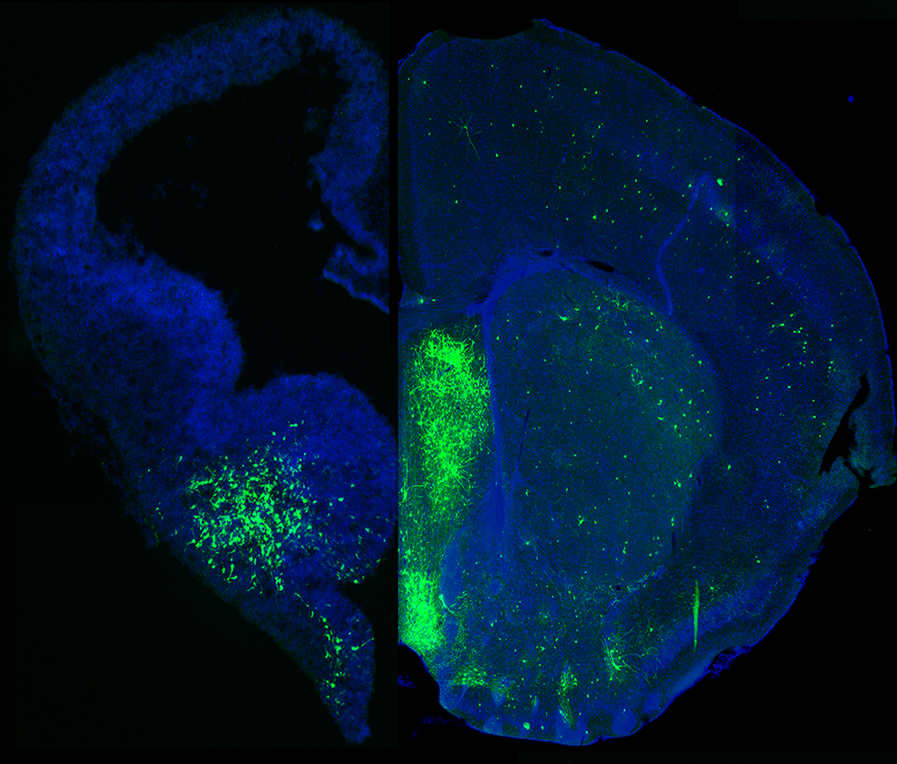
Figure 1. MGE–derived GABAergic cells populate many different brain regions.
The image depicts a section of an embryonic brain (left) that has been electroplated to label cells derived from the medial ganglionic eminence (MGE), merged with an section of an adult brain (right), displaying the incredible spatial and morphological diversity of MGE–derived cells in the mature brain. Understanding how this heterogeneous population is generated from one embryonic brain structure is the focus of this laboratory.
Mechanisms regulating initial fate decisions within the medial ganglionic eminence
The medial ganglionic eminence (MGE) gives rise to the majority of forebrain interneurons, most notably the somatostatin- and parvalbumin-expressing (SST+ and PV+) subtypes, and some nNOS (neuronal nitric oxide synthetase)–expressing neurogliaform and ivy cells in the hippocampus. The MGE is a transient, dynamic structure that arises around E10 and bulges into the lateral ventricle over the next several days before dissipating towards the end of embryogenesis. Given that initial fate decisions are generated within the MGE, there has been much focus on identifying a logic for interneuron generation from this region. Previous experiments characterized both a spatial and temporal gradient within the MGE that regulates the initial fate decision to become either PV+ or SST+ interneurons. SST+ interneurons are preferentially born early in embryogenesis from the dorsoposterior MGE, whereas PV+ interneurons are born throughout embryogenesis with a bias of originating from the ventroanterior MGE. Our work discovered an additional mechanism regulating this fate decision: the mode of neurogenesis. Using in utero electroporations, we found that PV+ interneurons are preferentially born from basal progenitors (also known as intermediate progenitors), whereas SST+ interneurons arise more commonly from apical progenitors. We hope to build on this observation to discover how these distinct spatial, temporal, and neurogenic gradients coordinate to regulate initial fate decisions of MGE progenitors.
Click image to view.
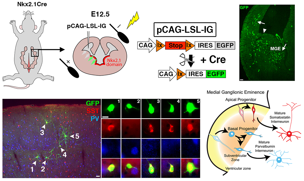
Figure 2. Manipulation of gene expression in the MGE by in utero electroporation (IUE)
Top. By using Nkx2.1-Cre mice, we can restrict expression of cre-dependent plasmids to the MGE. Note that the GFP+ cells are restricted to the MGE and to cells migrating away from the MGE at 2 days post-electroporation.
Bottom. Representative example from a P25 cortex of a mouse whose MGE was electroplated at E12.5. In this series of experiments, we used IUE to demonstrate that the mode of neurogenesis plays a role in interneuron fate determination: apical neurogenic divisions are biased to generate somatostatin (SST+) interneurons, whereas basal divisions primarily give rise to parvalbumin (PV+) cells.
Characterization of the epigenetic landscape during embryonic neurogenesis
In multicellular organisms, cells are genetically homogenous but structurally and functionally heterogenous as a result of differential gene expression that is often mitotically heritable. The mechanisms regulating such expression are 'epigenetic,' as they do not involve altering the DNA sequence itself; they include DNA methylation (DNAme), histone modifications, and higher-order chromatin structure. In particular, DNA and histone modifications often follow specific rules termed the “epigenetic code”, similar to the genetic code. Collectively, DNAme and histone modification have been reported to regulate transcription and chromatin structure in many stem cell and developmentally critical processes. Previous scRNA-seq (single-cell RNA sequencing) experiments on the ganglionic eminences (GEs) identified surprisingly few region-specific genes in cycling progenitors (immature cells that are still cycling and have not exited the cell cycle) despite the fact that these regions produce distinct GABAergic cell populations. Because there are dynamic changes in the chromatin landscape during development, a prevailing hypothesis is that epigenomic signatures may be a better predictor of cell fate during development, revealing both potential distal enhancers and/or genetic loci that may be ‘poised’ but not yet expressed. However, direct support for this hypothesis is lacking. This idea is particularly relevant, given that epigenetic changes are observed in many neurological and psychiatric diseases and that most single-nucleotide variants (SNVs) identified in diseases-specific GWAS (genome-wide association studies) map to non-coding regions, implying that epigenetic regulation of gene expression may underlie some disease etiologies. We are currently utilizing cutting-edge techniques to define the chromatin state of different embryonic neurogenic regions to better understand epigenetic changes in distinct cell types during development, with the hope applying this knowledge to various neurodevelopmental diseases.
Click image to view.
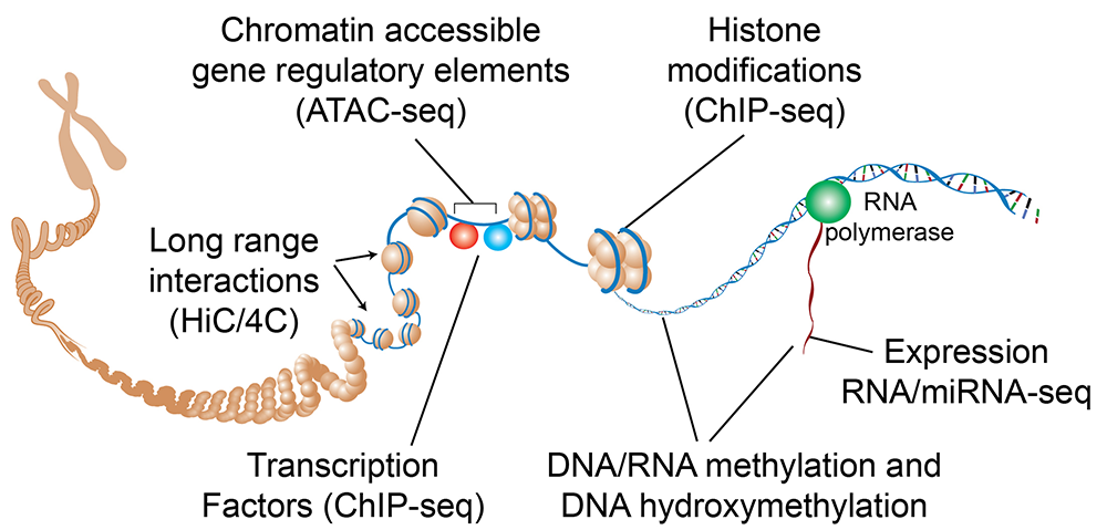
Figure 3. Schematic of epigenetic regulation mechanisms and applicable assays
While gene expression ultimately arises from RNA transcription, there are numerous epigenetic mechanisms that can regulate a cell's transcriptome, including DNA methylation, histone modifications, chromatin accessibility, and long-range chromatin interactions. There are numerous techniques available to assess all these distinct genomic DNA modifications. Image from Duke University Center for Genomic and Computational Biology.
How the environment sculpts interneuron diversity and maturation.
Interneurons undergo an extensive tangential migration period before reaching their terminal brain region, whereupon they interact with the local environment to differentiate and mature. The composition of interneuron subtypes varies significantly between different brain regions. Numerous experiments indicate that general interneuron classes, e.g., PV+– or SST+–expressing interneurons, are determined as cells become postmitotic during embryogenesis, but when other features that define a mature interneuron subtype (neurochemical markers, cell type, and subcellular location of synaptic partners, electrophysiology properties, etc.) are established remains unknown. One hypothesis is that interneurons undergo an initial differentiation into 'cardinal' classes during embryogenesis, and that maturation into 'definitive' subgroups requires active interaction with their mature environment. An alternate hypothesis is that immature interneurons are already genetically hardwired into definitive subgroups, and that the environment more passively sculpts the maturation of these cells. To test these competing hypotheses, we are harvesting early postnatal interneuron precursors (P0–P2) in specific brain regions and transplanting them into wild-type hosts either homotopically (cortex-to-cortex) or heterotopically (cortex-to-hippocampus or cortex-to-striatum). The technique allows us to determine whether transplanted interneurons adopt properties of the host environment (indicating a strong role for the environment in regulating interneuron diversity) or retain subtype features more consistent with the donor region. Our initial experiments indicate that the environment largely determines the composition of interneuron subtypes in a brain region regardless of donor region. However, some interneuron subtypes appear to be more genetically predefined and resistant to environmental influences than others. We are currently following up on these studies using scRNA-seq to characterize, in an unbiased manner, how a cell's transcriptome is altered when grafted into a new brain environment.
Click image to view.
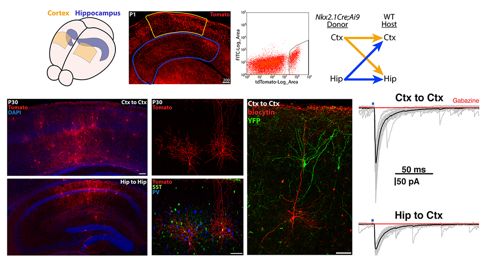
Figure 4. Transplantation of MGE–derived interneuron precursors into postnatal brains
Top. MGE–derived interneuron precursors are harvested from the cortex and hippocampus of P1 Nkx2.1-Cre;Ai9 mice, FACS–purified, and transplanted either homotopically (Ctx-to-Ctx, Hip-to-Hip) or heterotopically (Ctx-to-Hip, Hip-to-Ctx) into P1 wild-type (WT) mice.
Bottom. 30 days post-transplantation, tomato+ cells are dispersed throughout the host regions, displaying morphologies and neurochemical markers similar to endogenous interneurons. Grafted interneurons integrate into the host circuitry, as indicated by the postsynaptic responses in pyramidal cells upon stimulation of adjacent Nkx2.1-Cre;Ai32–derived, channel rhodopsin–expressing interneurons. Ctx: cortex; Hip: hippocampus.
Novel approach to identify genetic cascades underlying interneuron fate decisions
The ability to longitudinally track gene expression within defined populations is essential for understanding how changes in expression mediate both development and plasticity. Previous screens that were designed to identify genes and transcription factors specific to SST– or PV–fated interneurons were largely unsuccessful because several issues significantly hinder these types of studies. First, these interneurons originate from the MGE, which is a heterogeneous population of progenitors that give rise to both interneurons and a variety of GABAergic projection neurons, making it difficult to segregate interneuron progenitors from other cell types. Additionally, many markers that define mature interneuron subtypes are not expressed embryonically, and thus the class-defining markers are not helpful for studying MGE progenitors. In an ideal scenario, we would like to identify actively transcribed genes in MGE progenitors undergoing fate decisions while retaining the capacity to identify whether these cells become PV– or SST–expressing interneurons in the postnatal brain. To this end, we are developing a spatially and temporally inducible form of DNA adenine methylase identification (DamID), which will allow us to label the transcriptome of MGE progenitors. Labeled cells can be harvested at maturity, once we have the tools to distinguish between specific interneuron cell types. Then, the methylated genomic DNA will be analyzed, allowing us to look back in time to identify candidate fate-determining genes expressed in specific interneuron populations. Our hope is that the strategy could be widely applicable so that an investigator could characterize the temporal gene expression pattern of the cell type of interest.
Click image to view.
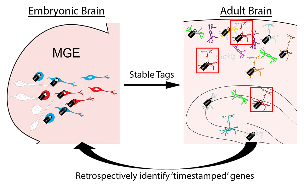
Figure 5. Timestamp of actively transcribed genes during development for future analysis
The goal of this approach is to label actively transcribed genes with stable methylation tags during embryogenesis as progenitors are undergoing initial fate decisions in the MGE. Then we can harvest specific interneuron subtypes in the adult brain using various transgenic mouse lines. Retrospective identification of an actively transcribed gene during embryogenesis will provide us with candidate fate-determining genes for specific interneuron subtypes.
Publications
- Quattrocolo G, Isaac M, Zhang Y, Petros TJ. Homochronic transplantation of interneuron precursors into early postnatal mouse brains. J Vis Exp 2018;136:e57723.
- Petros TJ. Stranger in a Strange Land: using heterotypic transplantations to study nature vs nurture in brain development. J Exp Neurosci 2018;12:1-4.
- McKenzie MG, Cobbs LV, Dummer PD, Petros TJ, Halford MM, Stacker SA, Zou Y, Fishell GJ, Au E. Non-canonical Wnt signaling through Ryk regulates the generation of somatostatin- and parvalbumin-expressing cortical interneurons. Neuron 2019;103(5):853-864.
- Wester JC, Madadevan V, Rhodes CT, Calvigioni D, Venkatesh S, Maric D, Hunt S, Yuan X, Zhang Y, Petros TJ, McBain CJ. Neocortical projection neurons instruct inhibitory interneuron circuit development in a lineage-dependent manner. Neuron 2019;102(5):960-975.
Collaborators
- Susan Amara, PhD, Laboratory of Molecular and Cellular Neurobiology, NIMH, Bethesda, MD
- Tudor Badea, MD, PhD, Retinal Circuit Development and Genetics Unit, NEI, Bethesda, MD
- Andrea Brand, PhD, The Gurdon Institute, University of Cambridge, United Kingdom
- Ryan Dale, PhD, Bioinformatics Core, NICHD, Bethesda, MD
- Dragan Maric, PhD, Flow and Imaging Cytometry Core Facility, NINDS, Bethesda, MD
- Chris McBain, PhD, Section on Cellular and Synaptic Physiology, NICHD, Bethesda, MD
- Isabel Perez-Otano, PhD, Instituto de Neurociencias de Alicante, Alicante, Spain
Contact
For more information, email tim.petros@nih.gov or visit https://www.nichd.nih.gov/research/atNICHD/Investigators/petros.