Immune Activation and Viral Pathogenesis

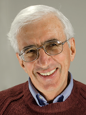
- Leonid Margolis, PhD, Head, Section on Intercellular Interactions
- Anush Arakelyan, PhD, Staff Scientist
- Christophe Vanpouille, PhD, Staff Scientist
- Rogers A. Ñahui-Palomino, PhD, Visiting Fellow
- Wendy Fitzgerald, BS, Biologist
The general goal of the Section for Intercellular Interactions is to understand the tissue-pathogenic mechanisms of human pathogens and the role of extracellular vesicles in such processes. We focused on the pathogenesis of the human immunodeficiency virus 1 (HIV-1) and on its co-pathogens. We found that extracellular vesicles (EVs) facilitate residual immune activation of HIV–infected human lymphoid tissue after viral replication is suppressed by antiviral therapy. In contrast, EVs released by vaginal microbiota bind to HIV-1 virions, preventing viral transmission. Thus, EVs play an important role in HIV infection and may serve a new target for therapy. Also, we also continue a project on SARS-CoV-2 pathogenesis.
Extracellular vesicles support residual HIV–triggered immune activation after HIV suppression.
Improper immune activation accompanies many human pathologies, including mental diseases, abnormal pregnancies, and HIV disease. In particular, immune activation is considered to be the driving force of HIV-1 disease, resulting in premature age-related diseases, such as cardiovascular disease or dementia. HIV–triggered immune activation continues for years, even when HIV-1 replication has been successfully suppressed by antiviral therapy (ART). Neither the causes nor the mechanisms of persistent systemic immune activation in HIV-1–infected patients under antiretroviral therapy are known. However, the residual immune activation is a major contributor to the early development of several pathologies in such patients. We addressed this problem in an ex vivo system of human lymphoid tissues that faithfully reflects many aspects of pathogenesis of various viruses in vivo. The goal of our work was to investigate the mechanisms of sustained immune activation in this system. Upon HIV infection, the ex vivo tissues become immuno-stimulated, faithfully reflecting the in vivo infection. We treated the infected tissues with several of the antivirals used for patients, in particular the protease inhibitor ritonavir (RTV) and the nucleoside reverse transcriptase inhibitors (NRTIs) zidovudine (AZT), in combination with lamivudine (3TC) and nevirapine (NVP, nonNRTI). All these compounds completely suppressed HIV replication. However, despite viral suppression, the tissues remained immune-activated, again reflecting the in vivo situation with ART–treated HIV–infected individuals. To understand the mechanisms underlying this phenomenon, we tested the following working hypotheses in this system.
- Immune activation is the result of the pro-inflammatory effects of certain antiretroviral drugs themselves. To test this, we compared cytokines released by ex vivo human lymphoid tissues under ART with those released by donor-matched untreated controls. However, ART did not lead to a significant increase in cytokines throughout the culture period.
- It is conceivable that an HIV–triggered upregulation of cytokines could stimulate immune cells to produce a long-term cascade of other cytokines, even after HIV-1 is suppressed. To investigate this possibility, we simulated this situation by treating uninfected ex vivo lymphoid tissues with a cocktail of cytokines that are upregulated in HIV-1 infection. However, tissue exposure to an exogenous cytokine combination did not result in a significant and sustained increase of cytokines.
- HIV-1 infection reactivates endogenous viruses, in particular human herpes viruses (HHVs), which may continue to replicate and induce immune activation after HIV is suppressed. To test this hypothesis, we quantified, by droplet digital PCR, HHVs 2–7 in tissues infected with HIV-1 and treated with ART. The only HHV that was upregulated upon HIV infection was cytomegalovirus. However, the upregulation was no longer significant after ART was applied.
- HIV-1 proteins that continue to be released, in spite of ART, activate the immune system. To test this hypothesis we treated ex vivo tissues with gp120, Tat, or Nef at a concentration that is comparable to that present in the viral inoculum used in our experiments. However, none of these tested HIV–encoded proteins induced any significant sustained cytokine upregulation.
Thus, our results did not support any of the four hypotheses. We therefore went on to propose that defective HIV-1 virions or extracellular vesicles carrying HIV components may trigger immune activation. We treated HIV-1 with aldrathiol-2 (AT-2), a compound that inactivates the virus but preserves virion morphology and its ability to interact with cell receptors. We inoculated tissues with AT-2–inactivated virions in the same amount as infectious virus. Repeated AT-2 HIV-1 exposure of tissues significantly upregulated cytokines. Even a single exposure of tissue to AT-2–inactivated HIV triggered sustained, significant immune activation.
We also applied EVs isolated from tonsil supernatants to the tissues. We found that EVs from tissues productively infected with HIV-1, and also from tissues in which HIV-1 replication was suppressed by ART, induced significant, sustained upregulation of cytokines. EVs isolated from control uninfected tissues or from uninfected tissues treated with ART did not produce a significantly sustained increase in cytokines compared with control untreated tissues.
In conclusion, the ex vivo lymphoid tissue system allowed the investigation, under laboratory-controlled conditions, of possible mechanisms involved in persistent immune activation in HIV-1 patients under ART. Our results with lymphoid tissue indicate that the mechanisms of sustained immune activation in these patients may include the presence of defective (replication-incompetent) virions and of EVs. These elements constitute potential therapeutic targets to combat the progression of various pathologies in HIV–infected individuals after HIV-1 itself has been successfully suppressed.
Vaginal bacteria–released extracellular vesicles in HIV infection
The vaginal microbiota of healthy reproductive-age women is generally dominated by Lactobacillus species. Lactobacilli are health-promoting microorganisms as they are involved in maintaining vaginal homeostasis by preventing overgrowth of pathogenic and opportunistic organisms. In particular, Lactobacilli have been reported to protect against vaginal transmission of HIV, although the mechanisms of protection remain unclear. Earlier, we established an ex vivo system of human cervico-vaginal tissue culture that recapitulates the features of HIV transmission and found in the system that several strains of Lactobacillus inhibited HIV-1 transmission.
We continued this work by investigating the mechanisms of this phenomenon, in particular the role of extracellular vesicles (EVs). EVs are released by both gram-negative and gram-positive bacteria. Like mammalian cells, bacterial EVs contain components from their mother cells and, despite a huge difference between bacterial and mammalian cells in size, structure, metabolism, and general physiology, the EVs that bacteria release are essentially of the same size. They carry diverse bioactive molecules, including proteins, nucleic acids, lipids, and metabolites. Although once thought to be useless cell debris, it is now clear that bacterial EVs are major players in important aspects of bacterial virulence, host immunomodulation, communication with other cells, survival, and other phenomena. Bacterial EVs have been implicated in bacteria-bacteria and bacteria-host interactions, promoting health or causing various pathologies.
After demonstrating for the first time that vaginal Lactobacilli isolated from the vaginas of healthy women (L. crispatus, L. gasseri) released nano-sized EVs similar to those released by Lactobacillus strains of gastrointestinal origin, such as L. casei, L. rhamnosus, L. reuteri, and L. plantarum, we investigated whether Lactobacillus-derived EVs are capable of inhibiting HIV-1 infection. Lymphocytic cell lines as well as human cervico-vaginal and tonsillar tissues ex vivo were infected with HIV-1 and treated with EVs from four different strains of Lactobacilli isolated from the vagina of healthy women. The choice of these Lactobacillus strains (L. crispatus BC3, L. crispatus BC5, L. gasseri BC12, and L. gasseri BC13) was based on our previous report on the anti-HIV-1 activity of these bacteria in human tissues ex vivo.
We found that EVs released by L. crispatus BC3 and L. gasseri BC12 largely protected human tissues ex vivo and T cells from HIV-1 infection. The HIV-1–inhibitory effects of EVs from L. crispatus BC3 or L. gasseri BC12 were dose-dependent. At the highest concentration used in our study there were about 1000 EVs per HIV-1 target cell. At this concentration, EVs were not cytotoxic, as evaluated with three different techniques (propidium-iodide-based assay, flow cytometry, and the MTT assay, which assesses cell viability). Thus, inhibition of HIV-1 infection was not the result of EV–induced cell death.
Not all Lactobacilli-released EVs inhibited HIV-1 infection: EVs from L. crispatus BC5 or L. gasseri BC13 did not. The inhibitory activity of EVs from L. crispatus BC3 and L. gasseri BC12 was therefore related to their composition, as the same number of EVs were used for all strains. Proteomic analysis showed that EVs that inhibited HIV-1 replication differ from those that did not, in terms of several proteins, namely enolase 2, 60 kDa chaperonin, elongation factor Tu, ATP synthase gamma chain, foldase protein PrsA 1, ATP synthase subunit delta, and triosephosphate isomerase. We identified several bioactive molecules in HIV-1–inhibiting EVs, in particular several enolases derived from Lactobacillus that were shown to inhibit the adherence of Neisseria gonorrhoeae to epithelial cells; also, bifidobacterial enolase, a cell-surface receptor for human plasminogen, was involved in the interaction with human host cells; the elongation factor Tu was shown to play an important role in the attachment of Lactobacillus johnsonii to human intestinal cells and mucins.
Whether the inhibition of HIV-1 entry is the result of the action of one or of a combination of several of these bioactive molecules acting synergistically remains an open question. Also, they may act only when associated with EVs. For these reasons, we next tested the effect of EVs as a whole in a cellular model of HIV-1 entry using TZM-bl cells. TZM-bl cells contain integrated reporter genes for firefly luciferase and E. coli β–galactosidase under the control of an HIV-1 long-terminal repeat, permitting sensitive and accurate measurements of infection at the entry/attachment level. We showed that viral attachment/entry to TZM-bl cells was inversely proportional to the concentration of bacterial EVs. Another set of experiments performed in the T cell line MT-4 confirmed that EVs directly inhibit HIV-1 attachment to cells. Thus, the anti–HIV-1 effect of Lactobacillus-derived EVs is mediated by the reduction in viral entry/attachment to the target cells, which appears to be related to direct alteration of HIV-1 virions by bacterial EVs. We found that virions pretreated with EVs released by L. crispatus BC3 and L. gasseri BC12, but not by L. crispatus BC5 or L. gasseri BC13, were no longer recognized by PG9, an antibody that specifically binds to functional trimeric gp120. Bacterial EVs from L. crispatus BC3 and L. gasseri BC12 interfere with the accessibility of viral Env, thus explaining the HIV-1 inhibition observed in cell lines and in human tissues ex vivo.
In summary, pretreatment of cells with bacterial EVs did not affect HIV-1 infection. However one treats cells with EVs and remove the vesicles, cells are as ineffaceable by the intact virus, as are control cells, i.e., viruses become defective and do not infect cells. Thus, bacterial EV–mediated HIV-1 inhibition is the consequence of EVs affecting the infectivity of virions rather than cell functions. In other words, of two participants of the infection, cells and viruses, EVs affect the latter. If confirmed in vivo, the finding may lead to new strategies to prevent male-to-female sexual HIV-1 transmission, for example by use of EVs derived from symbiotic bacteria.
SARS CoV-2 pathogenesis ex vivo
We continued our project on SARS-CoV-2 approved by the NIH Committee.
Investigating the mechanisms of SARS-CoV-2 tissue pathogenesis in vivo requires the development of an adequate system of human tissue culture under laboratory-controlled conditions. We developed such a system. Specifically, blocks of human lung tissue are cultured at the air-liquid interface on collagen sponges, and their cytoarchitecture is maintained for 14 days. Flow cytometry of cells from these blocks confirmed the viability of several cell types, including macrophages and other leukocytes, endothelial cells, and epithelial cells, and it also confirmed that the expression of the ACE-2 receptor, which the virus uses to invade a cell, is maintained. Histology revealed well preserved structural elements. Inoculation of these blocks with SARS-CoV-2 resulted in sustained viral replication and viral release into the culture medium. Flow cytometry identified infected cells, a finding that will be confirmed by immunohistochemistry. Analysis of culture medium from SARS-CoV-2–infected lung explants by multiplexed bead-based assays reveals that many cytokines are upregulated upon infection. The majority of the up-regulated cytokines are the same as those up-regulated in vivo.
Also, we used retroviral-based SARS-CoV-2 pseudoviruses with GFP and RFP protein reporters to study SARS-CoV-2 viral entry into target cells. Pseudoviruses were designed to express the SARS-CoV-2 spike (S) protein as well as other viral structural proteins, such as nucleocapsid (N), envelope (E), and membrane (M), in different combinations. Such one-cycle viruses infected 293T cell lines expressing ACE-2, and entry could be inhibited with neutralizing antibodies against the S protein. We found that M, N, and E proteins did not significantly affect the ability of the viruses to enter cells. In contrast, mutations in S protein that were identified in vivo changed the efficiency of pseudoviruses to enter cells. The United Kingdom B.1.1.7 variant infected 2.6 times more cells than pseudoviruses with wild-type S protein, and the South African variant B.1.351 infected 1.6 times more cells. Competition experiments between the pseudovariants is in progress and will reveal more information about how mutations in the S protein can affect entry in the setting of mixed viral populations.
To address pseudovirus binding to ACE-2 and to anti-S antibodies, we also designed a cell-free system. Using the magnetic nanoparticle (MNP) system that we had developed for analysis of HIV virions, we bound ACE2-Fc (Fc domain of human IgG linked to ACE-2) or anti-S antibodies to MNPs and evaluated the ability of pseudoviruses with S mutations to bind. The studies provide an alternate method of evaluating S mutations.
Furthermore, we demonstrated that primary human trophoblasts, isolated from placenta and shown to express ACE-2 and TMPRSS2, are susceptible to entry of SARS-CoV-2 pseudoviruses. We showed that the virus is able to enter the trophoblasts, as measured by presence of p24 retrovirus core protein inside the cells. However, the GFP reporter is rarely seen in these cells, indicating that restriction factors present in the trophoblasts prevent SARS-CoV-2 infection, implying that the placental anti-viral defense against SARS-CoV-2 likely involves post-entry processing.
In summary, all three systems, SARS-CoV-2–infected lung tissue ex vivo, pseudovirus cell infection of cells, and virus binding to nanoparticles-coupled SARS-CoV-2 cell receptors, can be used to study different aspects of viral pathogenesis and can be transformed into a platform to evaluate the efficiency of viral entry with S protein mutations, as well as for testing potential antivirals.
Additional Funding
- Office of AIDS Research (OAR), NIH, Intramural Award
Publications
- Ñahui-Palomino RA, Vanpouille C, Costantini PE, Margolis L. Microbiota–host communications: bacterial extracellular vesicles as a common language. PLoS Pathog 2021;17(5):e1009508.
- Mercurio V, Fitzgerald W, Molodtsov I, Margolis L. Persistent immune activation in HIV-1-infected ex vivo model tissues subjected to antiretroviral therapy: soluble and extracellular vesicle-associated cytokines. J Acquir Immune Defic Syndr 2020;84:45.
- Bhatti G, Romero R, Rice GE, Fitzgerald W, Pacora P, Gomez-Lopez N, Kavdia M, Tarca AL, Margolis L. Compartmentalized profiling of amniotic fluid cytokines in women with preterm labor. PLoS One 2020;15:e0227881.
- Sadovsky Y, Ouyang Y, Powell JS, Li H, Mouillet J-F, Morelli AE, Sorkin A, Margolis L. Placental small extracellular vesicles: current questions and investigative opportunities. Placenta 2020;102:34–38.
- Sass D, Saligan L, Fitzgerald W, Berger AM, Torres I, Barb JJ, Kupzyk K, Margolis L. Extracellular vesicle associated and soluble immune marker profiles of psychoneurological symptom clusters in men with prostate cancer: an exploratory study. Translat Psychiatry 2021;11:440.
Collaborators
- Michael Bukrinsky, MD, PhD, George Washington University, Washington, DC
- Leonid Chernomordik, PhD, Section on Membrane Biology, NICHD, Bethesda, MD
- Sara Gianella Weibel, MD, University of California San Diego, La Jolla, CA
- Michael Lederman, MD, Case Western University, Cleveland, OH
- Roberto Romero-Galue, MD, DMedSci, Perinatology Research Branch, NICHD, Detroit, MI
- Yoel Sadovsky, MD, Magee-Womens Research Institute, University of Pittsburgh, Pittsburgh, PA
- Alexandr Shpektor, MD, Moscow Medical University, Moscow, Russia
- Elena Vasilieva, MD, Moscow Medical University, Moscow, Russia
- Beatrice Vitali, PhD, Università di Bologna, Bologna, Italy
Contact
For more information, email margolis@helix.nih.gov or visit https://irp.nih.gov/pi/leonid-margolis.


