High-Resolution Structural Biology of Membrane Protein Complexes in Their Native Environment

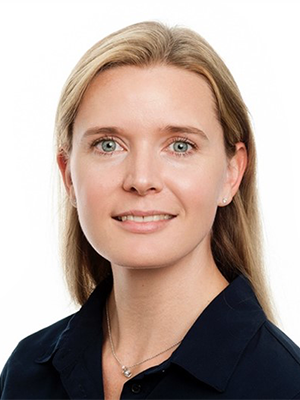
- Doreen Matthies, PhD, Head, Unit on Structural Biology
- Louis Tung Faat Lai, PhD, Visiting Postdoctoral Fellow
- Munazza Shahid, PhD, Visiting Postdoctoral Fellow
- Fei Zhou, PhD, Scientist
The Matthies lab is interested in the structure and function of membrane protein complexes in their native lipid membrane environment to understand their mechanism and the influence of their immediate surrounding and how these affect human health and disease. A cell contains many different lipid membranes with various lipid contents and distributions, which are very important for a membrane’s morphology and function. However, very little is understood about how these various micro-environments are formed and maintained and how they influence the structure and function of membrane proteins. Studying membrane protein complexes in their native biological membrane is therefore required.
We use a combination of molecular biology, biochemistry, and biophysical methods to study molecular transport across membranes with a focus on how the immediate native environment influences the structure and function of membrane proteins, but also how proteins and lipids shape and functionalize a lipid membrane.
Cryo-electron microscopy (Cryo-EM) is one of the main structural-biology methods of the lab. Using single-particle Cryo-EM, we solve high-resolution structures of membrane proteins in artificial environments such as in detergent micelles and lipid nano-discs. But we are extending this approach to studying membrane proteins in their native environment, using native lipid nano-discs, membrane fractions in forms of vesicles, and intact cells and tissues, using a combination of correlative light and electron microscopy techniques, including cryo-fluorescent microscopy, cryo-focused ion beam-scanning electron microscopy (Cryo-FIBSEM), Cryo-EM, and cryo-electron tomography (Cryo-ET).
Structural and functional investigation of chemokine receptors in their role in recurrent miscarriage
Recurrent miscarriage (RM) is usually defined as the loss of three or more consecutive pregnancies prior to the 20th week of gestation and affects approximately 1% of women of reproductive age. The cause of 50% of cases of RM is unknown, but there is evidence supporting immune causes, more specifically, that a T helper (Th) 1–type response is associated with the pathogenesis of RM. Women who suffer RM show elevated ratios of Th1 (CXCR3 and CCR5) to Th2 (CCR3 and CCR4) chemokine receptors. A Th1–type reaction in the materno-fetal interface mainly triggers an inflammatory response, while a Th2–type reaction typically promotes growth of trophoblastic cells, which is beneficial for the successful maintenance of a pregnancy. To work towards a treatment to prevent pregnancy loss in women with RM, Munazza Shahid will study the structure and function of chemokine receptors alone and in complex with their ligands and other interaction partners.
Structure and function of magnesium channels
Magnesium (Mg2+) is the most abundant divalent cation inside cells, with an average Mg2+ concentration of about 20 mM, most of it bound to proteins and ATP. Magnesium plays an essential role in cellular physiology, acting as a cofactor for more than 600 enzymes, including protein kinases, ATPase, exonucleases, and other nucleotide-related enzymes. Deficiency in Mg2+ is associated with such diseases as muscular dysfunction, bone wasting, immunodeficiency, cardiac syndromes, and neuronal disorders. The bacterial magnesium channel CorA is a homo-pentameric channel, which forms a symmetric closed state at normal to high concentrations of magnesium, with magnesium-binding sites between protomers as well as near the membrane pore. At low magnesium concentrations, the channel undergoes an asymmetric opening, which is likely to be caused by the destabilization of protomer interactions when magnesium ions dissociate from their binding site. Louis Lai will expand the research on magnesium channels, including looking at eukaryotic magnesium channels. To investigate the structure and mechanism of these channels, structural studies in synthetic as well as native nano-discs as well as in liposomes are planned.
Structural determination of the full-length SARS-CoV-2 spike protein and drug development
COVID-19 caused by the SARS-CoV-2 virus has posed a global threat since it was first identified at the end of 2019. The rapid development of vaccines has helped counteract the rapid spread of COVID-19. However, vaccines for children under the age of 12 have only just been approved. More children have been infected by more contagious variants of the virus, and the recent surge in COVID-19 cases has put an unprecedented pressure on the pediatric health care system. The SARS-CoV-2 spike protein is responsible for the initial binding of the virus to the ACE2 receptor on human cells. Better understanding of the function and structure of the spike protein is critical for primary prevention and for the development of a vaccine and therapeutic treatments to combat the COVID-19 pandemic. Structures of the spike protein’s soluble ectodomain have been determined, but the full-length spike, including its membrane domain, has not been well studied. We are working towards determining the structures of full-length spike protein complexes and identifying the key vaccine- and drug-binding interfaces in order to develop treatments that block viral entry into human cells with high efficiency and specificity, and which are also safe for children. Fei Zhou has successfully cloned and expressed the full-length spike protein, and we are working towards high-resolution structural determination of different variants and complexes.
Collaborations
Our collaborations involve structural and computational studies on a variety of membrane-protein complexes, including transporters, channels, and receptors, as well as viral spike proteins in different cellular compartments, virus-like-particles (VLP), SARS-CoV-2 accessory membrane proteins, extracellular vesicles, and lipid transport across cells, as well as novel detergents and polymers to gently extract membrane-protein complexes from their native lipid environment for high-resolution structural studies.
Publications
- He S, Chou H-T, Matthies D, Wunder T, Meyer MT, Atkinson N, Martinez-Sanchez A, Jeffrey PD, Port SA, Patena W, He G, Chen VK, Hughson FM, McCormick AJ, Mueller-Cajar O, Engel BD, Yu Z, Jonikas MC. The structural basis of Rubisco phase separation in the pyrenoid. Nat Plants 2020;6(12):1480–1490.
- Qiu B, Matthies D, Fortea E, Yu Z, Boudker O. Cryo-EM structures of excitatory amino acid transporter 3 visualize coupled substrate, sodium, and proton binding and transport. Sci Adv 2021;7:1–9.
Collaborators
- Nihal Altan-Bonnet, PhD, Laboratory of Host-Pathogen Dynamics, NHLBI, Bethesda, MD
- Tamir Gonen, PhD, Laboratory of Molecular Electron Microscopy, UCLA, Los Angeles, CA
- Rick K. Huang, PhD, NIH Intramural Research CryoEM Consortium, NCI, Bethesda, MD
- Maria S. Ioannou, PhD, Faculty of Medicine and Dentistry, University of Alberta, Edmonton, Canada
- Bechara Kachar, MD, Laboratory of Cell Biology, NIDCD, Bethesda, MD
- Gisela Storz, PhD, Section on Environmental Gene Regulation, NICHD, Bethesda, MD
- Sergei I. Sukharev, PhD, University of Maryland, College Park, MD
- Joshua J. Zimmerberg, MD, PhD, Section on Integrative Biophysics, NICHD, Bethesda, MD
- Manuela Zoonens, PhD, Institut de Biologie Physico-Chimique, Centre National de la Recherche Scientifique, Paris, France
Contact
For more information, email doreen.matthies@nih.gov or visit https://www.nichd.nih.gov/research/atNICHD/Investigators/matthies.


