Development of the Vertebrate Circulatory System

- Brant M. Weinstein, PhD, Head, Section on Vertebrate Organogenesis
- Aniket Gore, PhD, Staff Scientist
- Matthew Butler, PhD, Postdoctoral Fellow
- Hyun Min Jung, PhD, Postdoctoral Fellow
- Shlomo Krispin, PhD, Postdoctoral Fellow
- Mayumi Miller, PhD, Postdoctoral Fellow
- Timothy Mulligan, PhD, Postdoctoral Fellow
- Amber Stratman, PhD, Postdoctoral Fellow
- Jianxin Yu, PhD, Postdoctoral Fellow
- Olivia Farrelly, BS, Postbaccalaureate Fellow
- Kristin Johnson, BS, Postbaccalaureate Fellow
- Chase Melick, BS, Postbaccalaureate Fellow
- Van Pham, BS, Scientific Technician
The overall objective of this project is to understand how the elaborate networks of blood and lymphatic vessels arise during vertebrate embryogenesis. Blood vessels supply every tissue and organ with oxygen, nutrients, and cellular and humoral factors. Lymphatic vessels drain fluids and macromolecules from the interstitial spaces of tissues, returning them to the blood circulation, and play an important role in immune responses. Our studies on the formation of blood and lymphatic vessels are engendering intense clinical interest because of the roles both types of vessel play in cancer and ischemia. The zebrafish (Danio rerio) is a small tropical freshwater fish that possesses a unique combination of features that make it particularly suitable for studying vessel formation. Zebrafish are genetically tractable vertebrates with externally developing, optically clear embryos, which are readily available for observation and experimental manipulation. Such features make the fish highly advantageous for studying vascular development, permitting observation of every vessel in the living animal and simple, rapid screening for even subtle vascular-specific defects.
Current studies use genetic screening, experimental analysis, and imaging to examine cues directing vascular patterning and morphogenesis, regulation of vascular integrity, assembly of the lymphatic system, and hematopoietic stem cell formation.
Developing tools for experimental analysis of vascular development in the zebrafish
The development of new tools to facilitate vascular studies in the zebrafish is an important ongoing aim of our work. Previously, we (1) established a micro-angiographic method for imaging patent blood vessels in the zebrafish and used the method to compile a comprehensive staged atlas of the vascular anatomy of the developing fish (http://zfish.nichd.nih.gov); (2) generated a variety of transgenic zebrafish lines expressing various fluorescent proteins within vascular or lymphatic endothelial cells, making it possible for us to visualize vessel formation in intact, living embryos; and (3) developed methodologies for long-term multiphoton confocal time-lapse imaging of vascular development in transgenic fish. Recent technical advances have greatly facilitated the generation of new transgenic lines, and we are currently developing many new lines useful for in vivo vascular imaging as well as for in vivo endothelial-specific functional manipulation of signaling pathways involved in vascular specification, patterning, and morphogenesis. Furthermore, we are taking advantage of recently developed CRISPR (clustered regularly interspaced short palindromic repeats) gene-editing technology to easily generate germline mutations in any zebrafish gene of interest. We are also in the process of generating a detailed staged anatomical atlas of the developing lymphatic system in the zebrafish comparable to the atlas of the circulatory system that we had published previously.
Genetic analysis of vascular development
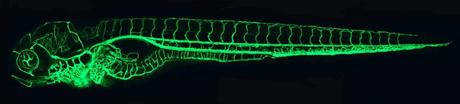
We use forward-genetic approaches to identify and characterize new zebrafish mutants that affect the formation of the developing vasculature. Using transgenic zebrafish expressing green fluorescent protein (GFP) in blood vessels (Figure 1) or lymphatic vessels, we are carrying out ongoing large-scale genetic screens for mutants induced by N-ethyl-N-nitrosourea (ENU). We already identified hundreds of new vascular mutants with phenotypes that include loss of most vessels or subsets of vessels, increased sprouting/branching, and vessel mis-patterning. We recently carried out a new genetic screen to identify hemorrhagic stroke susceptibility genes. We are currently using lymphatic-specific transgenic lines to perform screens for mutants specifically affecting the development of lymphatic vessels. We are pursuing the molecular cloning of the defective genes from all our new mutants, using next-generation whole-exome sequencing. To facilitate identification of the causative mutations (as opposed to naturally occurring polymorphisms) in our next-generation sequencing (NGS) data, we generated a large database of naturally occurring SNP (single nucleotide polymorphism) variants in three commonly used laboratory zebrafish strains. We also developed a web-based tool ("SNPfisher") to facilitate querying and manipulating the database. Whole-exome sequencing combined with our SNP identification tools has already made it possibly to rapidly identify the causative mutations in some of our more recently identified mutants. The identification of additional defective genes from our mutants should result in further new insights into the molecular mechanisms underlying vascular development and vascular integrity. Together, our ongoing mutant screens continue to yield a rich harvest of novel vascular mutants and genes, bringing to light new pathways that are critical during vascular development and vascular disease.
Click image to enlarge.
Figure 1. The zebrafish vascular system
Confocal microangiogram of the vascular system of a 4-1/2-day-old zebrafish larva labeled by injecting fluorescent microspheres. The transparency of zebrafish larvae makes it possible to use high-resolution optical imaging methods to visualize the entire vasculature in exquisite detail.
Analysis of vascular morphogenesis and integrity
Proper morphogenesis of vascular tubes and the maintenance of their integrity is of critical importance to human health. Malformation or rupture of vessels is the basis for stroke, the third leading cause of death and the most common cause of disability in developed nations. Intracerebral hemorrhage (ICH) accounts for 10 percent of stroke and is a particularly severe form of the disease, with disproportionately high rates of death and long-term disability. Therapeutic tools are still very limited, and prevention remains the most important way to reduce morbidity and mortality. In previous studies, we used high-resolution time-lapse two-photon imaging to examine vessel morphogenesis in living zebrafish, showing that the formation and intra- and intercellular fusion of endothelial vacuoles drive vascular lumen formation in vivo. We are currently examining the formation and maintenance of vascular endothelial cell-cell junctions, which are particularly important in stroke. To begin dissecting the molecular-regulatory mechanisms controlling endothelial junction formation, we examined a variety of genes required for vascular morphogenesis and vascular integrity, including the ccm, pak2a, and rap1b genes (see Figure 2 for the effects of rap1b disruption). We are also developing transgenic lines that permit us to visualize the dynamics of endothelial cell-cell junctions and intracellular cytoskeletal structures in order to examine their role in the cellular rearrangements that occur during vascular sprouting and growth and vascular tube formation. As noted above, we also carried out a genetic screen for genes that increase susceptibility to ICH. The screen identified many new mutants that elevate the propensity for ICH. Whole-exome NGS of the mutants is leading to the identification of novel genetic modifiers of the onset and severity of hemorrhagic stroke and, potentially, to the development of new therapeutic targets for the treatment and prevention of human ICH.
Click image to enlarge.
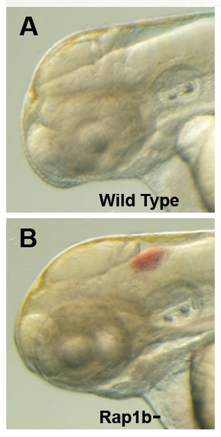
Figure 2. Intracranial hemorrhage (ICH) in the developing zebrafish
The clarity of zebrafish larvae also makes it straightforward to screen for animals with intracranial hemorrhage, as is evident in comparing lateral views of a 2-day-old wild-type larva (A) with a hemorrhage-prone larva deficient in rap1b (B).
Analysis of vascular patterning
We used multiphoton time-lapse imaging to characterize patterns of vessel assembly throughout the developing zebrafish. Our ongoing studies aim to understand how the patterns arise and what cues guide vascular network assembly during development. We previously demonstrated that known neuronal guidance factors play an important, previously unknown role in vascular guidance and vascular patterning, showing that semaphorin signaling is an essential determinant of trunk blood vessel patterning (Figure 3). More recently, we also showed that chemokine signaling orchestrates the assembly and patterning of the developing lymphatic vasculature of the trunk. Current studies are elucidating the role of additional factors that guide the patterning of developing blood and lymphatic vascular networks in vivo, both in the trunk and in vascular beds in the eye, aortic arches, hindbrain, and other anatomical locales.
Click image to enlarge.
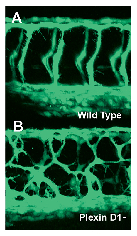
Figure 3. Mis-patterned trunk vessels in larvae lacking the vascular semaphorin receptor plexin D1
Confocal imaging of trunk vessels in a 2½-day-old wild-type (A) and a plexin D1–deficient (B) larva, showing loss of proper patterning of the trunk vessels caused by inability to receive semaphorin repulsive guidance signals.
Analysis of lymphatic development
The lymphatic system has become the subject of great interest in recent years because of the recognition of its important role in normal and pathological processes, but progress in understanding the origins and early development of the system has been hampered by difficulties in observing lymphatic cells in vivo and performing defined genetic and experimental manipulation of the lymphatic system in currently available model organisms. We recently demonstrated that the zebrafish possesses a lymphatic system that shares many of the morphological, molecular, and functional characteristics of the lymphatic vessels found in other vertebrates, thus providing a powerful new model for imaging and studying lymphatic development. As we continue to examine the origins and assembly of the lymphatic system of the zebrafish, we are developing new transgenic tools for imaging the development of the lymphatic system and for forward-genetic screening for lymphatic mutants. Our genetic analysis has already identified several novel genes involved in lymphatic development and patterning. We are also studying the roles of several different genes required for specification, assembly, or patterning of the lymphatic endothelium, including a role for chemokine signaling in guidance and patterning of lymphatic vessel assembly in the developing larval trunk. Our ongoing studies will thus provide new insights into the molecular regulation of lymphatic development.
Epigenetic regulation of hematopoietic stem and progenitor cell emergence
We recently discovered a novel mechanism for epigenetic regulation of hematopoietic stem and progenitor cell (HSPC) specification. HSPCs emerge from the ventral wall of the dorsal aorta in all vertebrates. We found that a gene encoding a DNA methyltransferase (dnmt) is expressed specifically in the ventral aortic endothelium, which gives rise to HSPCs. The gene functions downstream from a previously described genetic pathway for specification of HSPCs to promote the long-term maintenance of hematopoietic cell fate. Loss of the dnmt gene in vivo results in loss of HSPCs, while early ectopic overexpression of the dnmt gene is sufficient to induce ectopic hematopoietic gene expression. We are currently continuing to study the role of DNA methylation in HSPC formation.
Publications
- Butler M, Iben JR, Marsdan KC, Epstein JA, Granato M, Weinstein BM. SNPfisher: a tool for probing genetic variation in laboratory zebrafish. Development 2015; 142:1542-1545.
- Kallakuri S, Yu JA, Li J, Li Y, Weinstein BM, Nicoli S, Sun Z. Endothelial cilia are essential for developmental vascular integrity in zebrafish. J Am Soc Nephrol 2015; 26:864-875.
- Yu JA, Castranova D, Pham VN, Weinstein BM. Single cell analysis of endothelial morphogenesis in vivo. Development 2015; 142:2951-2961.
- Ulrich F, Carretero-Ortega J, Menéndez J, Sun B, Lancaster E, Pershad V, Trzaska S, Véliz E, Narvaez C, Kamei M, Prendergast A, Kidd KR, Shaw KM, Castranova DA, Pham VN, Lo BD, Martin BL, Raible DW, Weinstein BM, Torres-Va´zquez J. Reck enables cerebral vascular development by promoting Wnt/beta-catenin and VEGF signaling. Development 2016; 143: 147-159.
- Gore AV, Athans B, Iben JR, Johnson K, Russanova V, Castranova D, Pham VN, Iben JR, Butler MG, Williams-Simons L, Nichols JT, Bresciani E, Feldman B, Bresciani E, Kimmel C, Liu PP, Weinstein BM. Epigenetic regulation of hematopoiesis by DNA methylation. eLife 2015; 5.
Collaborators
- Maria Morasso, PhD, Laboratory of Skin Biology, NIAMS, Bethesda, MD
Contact
For more information, email weinsteb@mail.nih.gov.