About the Images
X-ray crystallography by Mitra Rana, a Visiting Fellow in the Banerjee Lab. 3D rendering by Jeremy Swan of the Computer Support Services Core.
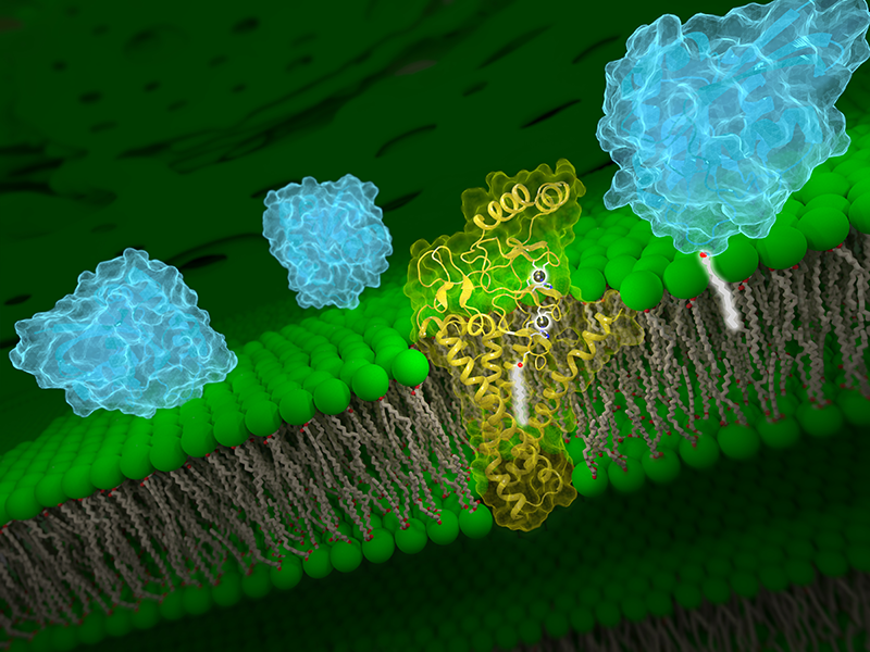
Molecular view of DHHC palmitoyltransferases. Human DHHC20 palmitoyltransferase (yellow) shown localized in the Golgi body membrane (green stacks). The Zn2+ ions are shown as gray spheres and the acyl chain of the palmitoyl group in white sticks. A hypothetical substrate (blue) approaches the palmitoyltransferase from the left and, after palmitoylation, is localized to the Golgi body membrane through anchoring of the palmitoyl group, now transferred to the substrate.
The molecular models were created with UCSF ChimeraX, developed by the Resource for Biocomputing, Visualization, and Informatics at the University of California, San Francisco, with support from National Institutes of Health R01-GM129325 and the Office of Cyber Infrastructure and Computational Biology, National Institute of Allergy and Infectious Diseases. The 3D render was created using Blender, a free, open-source 3D creation program.
Click image to view larger.