Neuregulin–ErbB Processing and Signaling in Neuronal Development and Psychiatric Disorders

- Andres Buonanno, PhD, Head, Section on Molecular Neurobiology
- Detlef Vullhorst, PhD, Staff Scientist
- Irina Karavanova, PhD, Research Assistant
- Tanveer Ahmed, PhD, Postdoctoral Fellow
- Lalitha Kurada, PhD, Postdoctoral Fellow
- Miguel Skirzewski, PhD, Postdoctoral Fellow
- Larissa Erben, MS, Graduate Student
- Marie Cronin, BS, Postbaccalaureate Fellow
Failure of cortical microcircuits to properly regulate excitatory/inhibitory (E/I) balance is a key feature in the etiology of several developmental psychiatric disorders and neurological diseases, such as schizophrenia, autism, ADHD, and epilepsy. E/I balance is important to synchronize the firing pattern of local neuron ensembles, and its dysregulation can degrade cognitive functions and, in extreme cases, result in epileptiform activity. Network activity, in particular oscillatory activity in the gamma-frequency range (30–80 Hz), is altered in many psychiatric disorders and may account for their cognitive and behavioral symptoms. We are interested in how Neuregulin and its receptor ErbB4, which are both genetically linked to psychiatric disorders, function in rodents in an activity-dependent fashion (i.e., experience) in the developing brain to regulate synaptic plasticity, neuronal network activity, and behaviors that model features of many psychiatric disorders. We identified functional interactions between the Neuregulin/ErbB4, dopaminergic, and glutamatergic signaling pathways in GABAergic interneurons that are critical for understanding how Neuregulins regulate neuronal plasticity, E/I balance, and synchronous activity in neuronal networks.
Our earlier studies demonstrated that in the hippocampus and neocortex expression of ErbB4, the major Neuregulin neuronal receptor, is restricted to GABAergic interneurons. ErbB4 levels are especially high in a subtype of GABAergic neurons, known as parvalbumin-positive (Pv+) fast-spiking interneurons, that are necessary for the generation and modulating of gamma oscillations. Using genetically targeted mouse mutant models, we went on to show that Neuregulin–ErbB4 signaling regulates synaptic plasticity, neuronal network activity (i.e., gamma oscillations), and behaviors associated with psychiatric disorders. Our group more recently investigated other aspects of Neuregulin brain expression: the subcellular expression and trafficking of different Neuregulin isotypes, their post-translational processing in response to neuronal activity, and their functions in the developing and maturing nervous system. To achieve these goals, we use a combination of techniques, including: electrophysiological recordings in acute brain slices prepared from normal and genetically altered mice; multi-electrode field recordings from brains of freely moving rats; reverse-microdialysis neurochemistry; confocal fluorescence microscopy in fixed and live tissue; proteomics analyses; and behavioral testing. The ultimate goal of this multi-disciplinary approach is to generate holistic models to investigate the developmental impact of genes that modulate E/I balance and neuronal network activity and that consequently affect behaviors and cognitive functions altered in many psychiatric disorders.
Subcellular distribution and functions of single transmembrane (TM) Neuregulins in central neurons
Numerous Neuregulins (NRGs) are generated through the use of four different genes (NRG1–NRG4), promoters (NRG1: types -I, -II and -III), and alternative splicing, but the functional significance of this evolutionarily conserved diversity remains poorly understood. The cellular and molecular processes that promote the conversion of NRG ligands from inactive pro-forms to signaling-competent ligands that can engage ErbB4 receptors to mediate their numerous biological effects in the developing and maturing brain remain mostly unknown. As discussed here and further below, our recent studies reveal that NRGs can be categorized by their distinct membrane topologies, which impart fundamentally different subcellular trafficking properties. NRGs whose pro-forms contain a single transmembrane domain, such as NRG1 (types I and II) and NRG2, target to the cell body plasma membrane, where they accumulate at specialized contact sites with the underlying endoplasmic reticulum. We began our studies by analyzing NRG2, an isotype that is prominently expressed in the developing postnatal and adult CNS. Using a novel double-labeling in situ hybridization technique (RNAScope) and newly generated monoclonal antibodies, we found that, in the rodent hippocampus, NRG2 mRNA and protein are highly expressed in ErbB4–positive GABAergic interneurons, suggesting that NRG2 and ErbB4 can engage in autocrine signaling in this neuron type. Interestingly, we found no evidence of NRG2 protein in axons or distal dendrites; instead, we found that unprocessed proNRG2 accumulates as large somato-dendritic puncta on the neuronal soma and proximal dendrites (Reference 2). Our more recent studies on the other single TM NRGs (NRG1 types I and II) demonstrate a similar subcellular distribution. Moreover, we found that the ectodomains of single TM NRGs are cleaved by sheddases in an activity-dependent manner to signal in paracrine and autocrine fashion, and that this activity is mediated by NMDA receptor activation on cortical interneurons to promote proNRG2 shedding and subsequent ErbB4 receptor signaling (Figure 1 A,B). In turn, the activation of ErbB4 promotes its association with NMDARs and their internalization. In this fashion, there is bidirectional signaling between NRG/ErbB4 and NMDAR that can function as a homeostatic mechanism to regulate interneuron excitability (References 1,2).
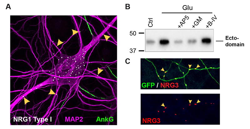
Click image to enlarge.
Figure 1. Differential trafficking of NRG isoforms in transfected hippocampal neurons
A. NRG1 type I accumulates as puncta on soma and proximal MAP2–positive dendrites, while axons (proximal segments identified by Ankyrin G) and more distal dendrites are negative. B. Stimulation of neurons with 20 μM glutamate (Glu) promotes NRG1 ectodomain shedding. The effect is blocked by NMDA receptor (AP5) or metalloprotease (GM6001) inhibition. C. NRG3 puncta accumulate on GFP–filled axons, away from cell bodies.
Subcellular distribution of dual transmembrane Neuregulins in central neurons
By contrast to single TM NRGs, we found that dual TM NRGs, such as CRD (cysteine-rich domain)–NRG1 (type III) and NRG3, for which we recently reported its unexpected dual TM topology (Reference 1), are targeted to axons where they signal in juxtacrine mode (Figure 1C). Moreover, unlike single TM NRGs, processing of dual TM NRGs is constitutive and independent of NMDA receptor activity. Interestingly, processing is a prerequisite for axonal accumulation of dual TM NRGs; treatments that interfere with processing cause intracellular accumulation of their pro-forms. Taken together, our recent findings about the relationships between transmembrane topology, modes of proteolytic processing, subcellular distribution, and signaling modality suggest that single- and dual-pass NRGs regulate neuronal functions in fundamentally different ways (Figure 1C,D). This work was supported by a Directors Investigator Award.
Neuregulin-2 knockout mice exhibit dopamine dysregulation and severe behavioral phenotypes with relevance to psychiatric disorders.
We found that NRG2 expression in the adult rodent brain does not overlap with NRG1 and is more extensive than originally reported, including expression in the striatum and medial prefrontal cortex (mPFC). We therefore generated NRG2 knockout (KO) mice to study NRG2's function (Figure 2). NRG2 KOs have higher extracellular dopamine levels in the dorsal striatum but lower levels in the mPFC than wild-type mice—a pattern with similarities to dopamine dysbalance in schizophrenia. Like ErbB4 KO mice, NRG2 KOs performed abnormally in a battery of behavioral tasks relevant to psychiatric disorders. NRG2 KOs exhibit hyperactivity in a novelty-induced open field, deficits in prepulse inhibition, hypersensitivity to amphetamine, antisocial behaviors, reduced anxiety-like behavior in the elevated plus maze and deficits in the T-maze alteration reward test—a task that is dependent on hippocampal and mPFC function. Acute administration of clozapine rapidly increased extracellular dopamine levels in the mPFC and improved alternation T-maze performance (Figure 2D). Similar to mice treated chronically with N-methyl-D-aspartate receptor (NMDAR) antagonists, we demonstrated that NMDAR synaptic currents in NRG2 KOs are augmented at hippocampal glutamatergic synapses and exhibit augmented sensitivy to ifenprodil, indicating an elevated contribution of GluN2B–containing NMDARs. Our findings reveal a novel role for NRG2 in the modulation of behaviors with relevance to psychiatric disorders (Reference 3).
Click image to enlarge.
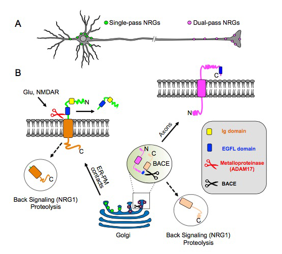
Figure 2. Transmembrane (TM) topology of unprocessed pro–NRGs determines their neuronal subcellular targeting and processing in central neurons.
A. A schematic presentation, based on our findings (Reference 1), that depicts single TM NRGs accumulating at somatic subsurface cistern and at proximal dendrites to mediate NRG\ErbB4 signaling, whereas dual TM NRGs accumulate on axons and their terminals to drive long-range signaling.
B. Single TM NRGs are trafficked through the secretory pathway as unprocessed pro-forms and accumulate at subsurface cisternae (SSC) sites, where NMDAR activity promotes ectodomain shedding. By contrast, unprocessed dual TM NRGs traffic via the Golgi and are cleaved by BACE (beta-secretase) to release the amino-terminal region, which is transported into axons in an anterograde manner. Both single– and dual–TM NRGs are processed by gamma-secretase to release carboxyl-terminal proteolytic fragments, which can “back-signal” into the nucleus to regulate transcription.
Analysis of ErbB4 function in mice harboring targeted mutations in GABAergic and dopaminergic neurons
Dysfunctional NRG/ErbB4 signaling in the hippocampus, pre-frontal cortex (PFC), and striatum may contribute to alterations in dopamine (DA) function associated with several schizophrenia symptoms. Given that we had shown that NRG1 acutely increases extracellular DA levels to regulate long-term potentiation (LTP) and gamma oscillations, and that ErbB4 receptor expression is confined to GABAergic interneurons (cortex) and TH+ mesocortical DAergic neurons, we used genetic, biochemical, and behavioral approaches to measure DA function in the hippocampus, PFC, and striatum in mice harboring targeted mutations of ErbB4 in either PV+ or TH+ neurons. Interestingly, we have found that, in contrast to GABAergic neurons, ErbB4 is highly expressed on the axons of DA neurons, suggesting that NRG/ErbB4 signaling may directly regulate the presynaptic function of these neurons. We found NRG regulates the increase in extracellular DA levels, at least in part, by regulating DA transporter (DAT) function. In contrast to mice harboring CNS–wide or GABAergic–restricted mutation of ErbB4, which show sensory-motor gating deficits and increases in motor activity, mice with ablation of ErbB4 in TH+ neurons only manifest behavioral deficits in cognitive-related tasks, such as performance on the T-maze, Y-maze, and Barnes maze. Our findings suggest that direct effects of NRG/ErbB4 signaling in GABAergic and DAergic neurons in combination regulate cortical circuits and DA homeostasis to affect numerous behaviors relevant to schizophrenia (Reference 4).
A novel exon-specific in situ hybridization approach uncovers a new pattern of ErbB4 isoform expression in brain neurons and oligodendrocytes.
ErbB4 receptor isoforms are generated by the alternatively splicing of exons that encode either one of the two juxtamembrane domains (JMa/JMb) and either one of the cytoplasmic domains (CYT-1/CYT-2), which impart different stability and downstream signaling modes to the receptor. Importantly, the ratios of JMa/JMb– and Cyt-1/Cyt-2–containing ErbB4 receptor isoforms are specifically augmented in the postmortem prefrontal cortex of individuals with schizophrenia. In order to study expression of these four alternatively spliced ErbB4 variants at a single-cell level, we implemented a novel highly sensitive fluorescent/colorimetric in situ hybridization approach that uses single-pair oligonucleotide probes (about 50bp) targeting exon/exon splice junctions. By comparing ErbB4 hybridization on sections from wild-type (WT) with ErbB4 knockout mice (missing exon 2), we initially demonstrated that single-pair probes provide the sensitivity and specificity to visualize and quantify the expression of ErbB4 isoforms (Figure 3A–F). Our analyses then identified brain regions of differential ErbB4 isoform expression conserved from mouse to humans. Using transgenic mice that drive the GFP reporter selectively in either neurons or glial cells, we went on to demonstrate that expression of distinct ErB4 isoforms differs between neurons and cells of the oligodendrocyte lineage. Interestingly, GABAergic interneurons mostly express the ErbB4 JMb isoform, whereas oligodendrocytes predominantly or exclusively express JMa receptor variants (Fig. 3G–J). Whereas the original reports attributed the changes of ErbB4 isoforms in postmortem brain to the result of altered expression in GABAergic neurons, our findings suggest that the increased JMa/JMb ratio observed in the postmortem brains of schizophrenia subjects could result from expression of ErbB4 JMa isoforms in cortical oligodendrocytes or their progenitors.
Click image to enlarge.
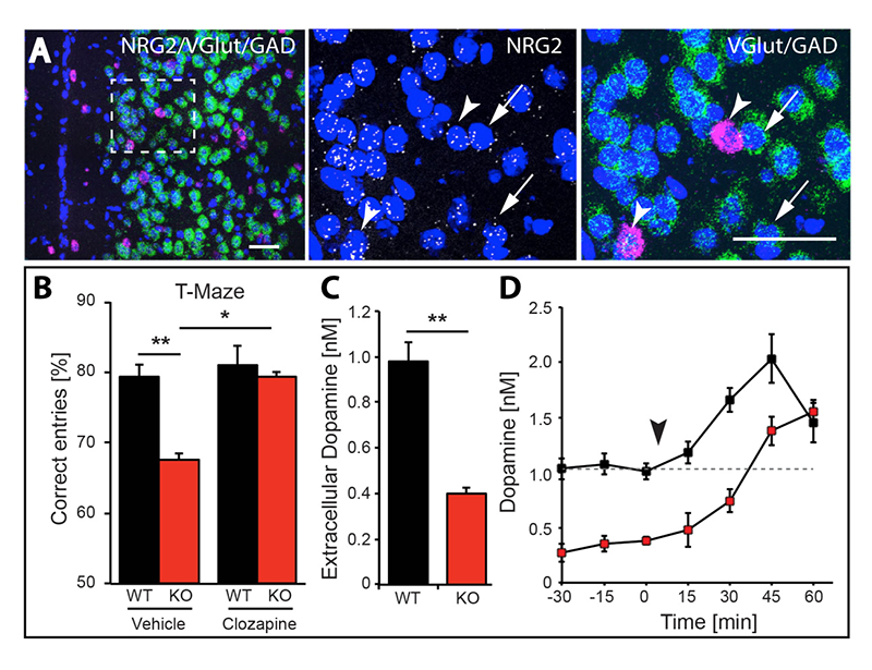
Figure 3. NRG2 null mice exhibit working memory deficits and reduced dopamine levels that are restored by clozapine.
A. Expression of NRG2 in the prefontal cortex (PFC) analyzed by triple in situ hybridization. NRG2 transcripts (white) are expressed in both glutamatergic (green) and GABAergic (magenta) neurons.
B. Poor performance by NRG2 knockout (KO) mice in a T-maze reward alternation task, as compared with WT littermates (left), can be restored by administration of the antipsychotic clozapine (right).
C. Reduced extracellular dopamine levels in the mPFC of NRG2 KO mice.
D. Extracellular dopamine levels in the mPFC of NRG2 KO mice rise after clozapine injection (arrowhead) at a time that coincides with improved performance on the T-maze.
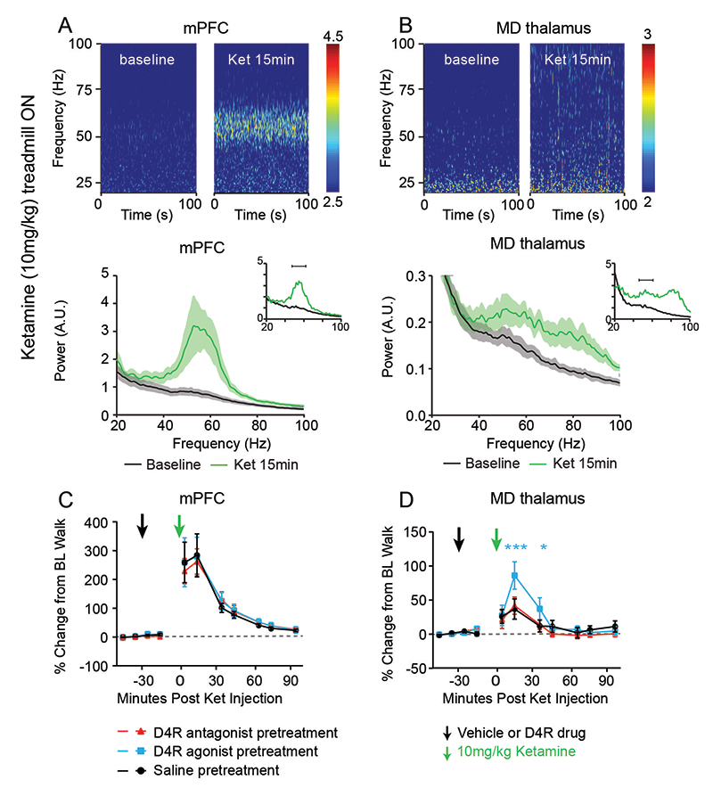
Effects of ketamine on cortical gamma oscillations and the role of dopamine receptors
Mounting evidence suggests that gamma oscillations are atypically high at baseline in disorders that affect attention, such as schizophrenia and ADHD. Ketamine, an antagonist of the NMDAR, has profound effects on gamma oscillation power, and it phenocopies schizophrenia by eliciting psychoto-mimetic symptoms and affecting cognitive functions in healthy individuals. In collaboration with Judith Walters’ lab, we used multi-electrode recordings from the medial prefrontal cortex (mPFC) and mediodorsal thalamus (MD) of rats acutely treated with ketamine, which serves as a rodent model with “face validity” for schizophrenia, to study the drug’s effects on spiking and gamma local field potentials in the mPFC and MD of freely moving rats. We found that ketamine raises gamma local field potentials and frequencies in both brain areas, but does not increase thalamocortical synchronization. Based on our prior in vitro studies, showing that a “cross-talk” between dopamine D4 (D4R) and ErbB4 receptors regulate gamma oscillation power in acute hippocampal slices, we investigated whether and how D4R–targeting drugs regulate gamma oscillations. We found that a D4R agonist (A-412997) increased ketamine-induced gamma power that was blocked by a D4R–selective antagonist (L-745870) in both mPFC and MD, but that neither drug altered ketamine-induced gamma power or frequency in the mPFC. Interestingly, in the MD, the D4R agonist increased the power of ketamine-induced gamma oscillations (Reference 5). Experiments are in progress to evaluate the effects of drugs targeting dopamine D4R receptors on attention and impulsivity behaviors in mice, using the five-choice serial-reaction time task (5CSRTT).
Click image to enlarge.
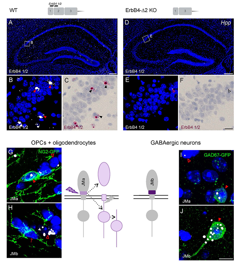
Figure 4. Oligodendrocytes and neurons express distinct ErbB4 JM transcripts.
The sensitivity and specificity of single-pair probe in situ hybridization was demonstrated by the presence of signals in sparse GABAergic hippocampal neurons of wild-type (WT) mice hybridized with a probe corresponding to exon 2 of the ErbB4 receptor gene (A–C), and by the absence of signal in sections prepared from ErbB4 knockout mice that lack exon 2 (D–F). Oligodendrocyte precursors cells (OPCs) and mature oligodendrocytes express ErbB4 JMa isoforms, which are susceptible to shedding and can back-signaling (G,H), whereas GABAergic neurons express the cleavage-resistant JMb ErbB4 receptor (I,J).
Click image to enlarge.
Figure 5. Ketamine-induced gamma oscillations in the medial prefrontal cortex (mPFC) and medial thalamus (MD) of walking rats, and regulation by D4R agonist
Multi-electrode recordings of local field potentials in the (A) mPFC and (B) MD reveal a dramatic increase in gamma frequency (40–70Hz) power in walking rats following ketamine injection (green), relative to baseline (BL) walking by rats injected with saline (black). C,D. Effects of D4R drugs on ketamine-induced gamma power. Administration of saline or a D4R antagonist (red) had no effect on ketamine-induced gamma power in either the (C) mPFC or the (D) MD, whereas the D4R agonist A-412997 (cyan) selectively increased gamma power in the MD.
Additional Funding
- Bench-to-Bedside Award (NHD15003-001-00001) “Selective Dopamine D4 Receptor-Targeting Compounds as Pro-Cognitive Drugs”
- NICHD DIR Director's Investigator Award (RRC# D-14-07). PI: Andres Buonanno; Co-PI: Juan Bonifacino. “Exploring the functional role of Neuregulin isoform diversity in CNS”
Publications
- Vullhorst D, Ahmad T, Karavanova I, Keating C, Buonanno A. Structural similarities between Neuregulin 1-3 isoforms determine their subcellular distribution and signaling mode in central neurons. J Neurosci 2017 37:5232-5249.
- Vullhorst D, Mitchell RM, Keating C, Roychowdhury S, Karavanova I, Tao-Cheng JH, Buonanno A. A negative feedback loop controls NMDA receptor function in cortical interneurons via neuregulin 2/ErbB4 signalling. Nat Commun 2015 6:7222.
- Yan L, Shamir A, Skirzewski M, Leiva-Salcedo E, Kwon OB, Karavanova I, Paredes D, Malkesman O, Bailey KR, Vullhorst D, Crawley JN, Buonanno A. Neuregulin-2 ablation results in dopamine dysregulation and severe behavioral phenotypes relevant to psychiatric disorders. Mol Psychiatry 2017; ePub ahead of print.
- Skirzewski M, Karavanova I, Shamir A, Erben L, Garcia-Olivares J, Shin JH, Vullhorst D, Alvarez VA, Amara SG, Buonanno A. ErbB4 signaling in dopaminergic axonal projections increases extracellular dopamine levels and regulates spatial/working memory behaviors. Mol Psychiatry 2017; ePub ahead of print.
- Furth KE, McCoy AJ, Dodge C, Walters JR, Buonanno A, Delaville C. Neuronal correlates of ketamine and walking induced gamma oscillations in the medial prefrontal cortex and mediodorsal thalamus. PLoS One 2017 12(11):e0186732.
Collaborators
- Veronica Alvarez, PhD, Section on Neuronal Structure, NIAAA, Rockville, MD
- Susan G. Amara, PhD, Laboratory of Molecular and Cellular Neurobiology, NIMH, Bethesda, MD
- Jung Hwa (Susan) Cheng, PhD, EM Facility, NINDS, Bethesda, MD
- Claire Delaville, PhD, Experimental Therapeutics Branch, NINDS, Bethesda, MD
- Katrina Furth, PhD, Experimental Therapeutics Branch, NINDS, Bethesda, MD
- Jennie Garcia-Olivares, PhD, Laboratory of Molecular and Cellular Neurobiology, NIMH, Bethesda, MD
- Idit Golani, MD, Technion–Israel Institute of Technology, Haifa, Israel
- Ilana Kremer, MD, Technion–Israel Institute of Technology, Haifa, Israel
- Sanford P. Markey, PhD, Laboratory of Neurotoxicology, NIMH, Bethesda, MD
- Alon Shamir, PhD, Mazra Mental Health Center, Akko, Israel
- Jung-Hoon Shin, PhD, Section on Neuronal Structure, NIAAA, Rockville, MD
- Judith R. Walters, PhD, Experimental Therapeutics Branch, NINDS, Bethesda, MD
Contact
For more information, email buonanno@mail.nih.gov or visit http://smn.nichd.nih.gov.