Deciphering Microbial Virulence Mechanisms

- Matthias Machner, PhD, Head, Unit on Microbial Pathogenesis
- Devanand Bondage, PhD, Visiting Fellow
- Eric Cheng, PhD, Visiting Fellow
- Nicole Ellis, PhD, Visiting Fellow
- Pei-Chung Lee, PhD, Visiting Fellow
- Xiao Li, PhD, Visiting Fellow
- Yi-han Lin, PhD, Visiting Fellow
- Anne Davidson, BS, Postbaccalaureate Student
- Katherine Gillis, BA, Postbaccalaureate Student
Our main research goal is to obtain mechanistic insight into the virulence strategies of microbial pathogens. As a model organism, we use the bacterium Legionella pneumophila, the causative agent of a potentially fatal respiratory infection known as Legionnaires' disease. Contrary to what its name may imply, Legionnaires’ disease occurs in individuals of all ages, including children who receive respiratory therapy, newborns who had recently undergone surgery or under-water birth, and children who are immune-compromised. We are committed to an in-depth analysis of mechanisms that allow L. pneumophila to exploit the human host and cause disease. Insights gained from these studies will ultimately improve our ability to diagnose, prevent, and fight Legionnaires’ disease and related illnesses, thereby contributing to the success of NICHD’s mission.
Upon inhalation of contaminated water droplets, L. pneumophila enters the lung and is phagocytosed (taken up) by alveolar macrophages, specialized immune cells. Instead of being degraded by these cells, the pathogen establishes a protective membrane compartment, the Legionella-containing vacuole (LCV). Within this intravacuolar niche, L. pneumophila can replicate to high numbers before killing the host cell and infecting neighboring cells.
Virulence of L. pneumophila relies on the activity of close to 300 proteins, or effectors, that are delivered into the host cytosol by a specialized translocation apparatus called Dot/Icm type IV secretion system (T4SS). L. pneumophila mutants with a non-functional type IV secretion system are degraded by macrophages, underscoring the importance of the translocated effectors for host cell manipulation and bacterial virulence.
Our main research objective is to obtain detailed mechanistic insight into the regulation and function of L. pneumophila effectors by investigating host-pathogen interactions at a molecular, cellular, and structural level. Deciphering the virulence program of this dangerous pathogen will set the stage for the development of novel therapeutics aimed at treating or preventing Legionnaires' disease and related illnesses.
A bacterial E3 ligase relic hijacks host-cell ubiquitination.
Bacterial pathogens often target conserved host pathways by encoding proteins that are molecular mimics of cellular enzymes, thus tricking the host cell into surrendering its resources to the bacteria. We discovered that L. pneumophila uses such a strategy to exploit ubiquitination, a conserved post-translational modification that is mediated by a family of enzymes called E3 ubiquitin ligases. L. pneumophila encodes its own molecular mimics of E3 ligases, including the effector protein RavN, thereby subverting the ubiquitin pathway for its own benefit during infection. By testing truncated RavN variants in an in vitro reconstitution assay, we found that the E3 ligase activity of RavN is located within its N-terminal region. Using protein crystallography, we revealed that the fold of RavN shows only residual resemblance to conventional eukaryotic E3s. The N-terminal region of RavN displays a U-box–like domain, a structural motif that mediates the interaction with E2 ubiquitin–conjugating enzymes. In RavN, the U-box lacks the central alpha helix commonly found in other U-box domains, indicating that RavN is an E3 ligase relic that has undergone significant evolutionary alteration. Yet its mode of interaction with E2 enzymes, host proteins that are important for the ubiquitin transfer reaction, has been preserved throughout evolution, and substitution of amino acid residues within the predicted E2 binding interface render RavN inactive.
Identification of catalytic activities in Legionella effectors
Using RavN as a model for an in silico analysis, we discovered several additional E3 ligase mimics within the effector repertoire of L. pneumophila that, similar to RavN, lack significant homology to known E3s but, nonetheless, catalyze the ubiquitination reaction. Our findings support the hypothesis that E3 ligases have been a vital part of the virulence program of L. pneumophila and that these effectors, despite having undergone extensive evolutionary changes, retain features that are critical for their biological function, including the ability to hijack factors that are part of the host ubiquitination machinery. The findings indicate that ubiquitination is more extensively exploited during infection by L. pneumophila than previously thought and that interference with this post-translational modification could constitute a novel therapeutic strategy to antagonize infections by L. pneumophila and related pathogens.
‘Smarter’ drugs that selectively target pathogenic bacteria
Most classical antibiotics kill bacteria or inhibit their growth by targeting key steps in their physiology. This approach, while effective in the past, has led to the rapid emergence of multidrug-resistant strains that have become insensitive to the microbicidal or microbiostatic activity of existing antibiotics. In addition, recent insight into the complexity of the human microbiome and its importance for human health has raised additional concerns about the excessive use of antibiotics and their collateral effect on the commensal microbiota. Thus, there is an urgent need for the development of ‘smarter’ therapeutics that discriminate between pathogens and commensals by selectively targeting virulence mechanisms of bacteria.
Given their essential role in virulence, the L. pneumophila T4SS and its translocated effectors represent compelling targets for the development of novel therapeutic agents. In collaboration with the National Center for the Advancement of Translational Sciences (NCATS), we screened over 18,000 bioactive compounds from various product libraries for their ability to target Legionella virulence factors. Candidate compounds that emerged from this screen underwent multiple rounds of rigorous validation and testing in order to select the most efficient molecules. Ultimately, we identified a small group of compounds that protected human macrophages from intracellular replication of L. pneumophila, making them ideal candidates for an in-depth analysis of their therapeutic potential. At the same time, these compounds are being analyzed in detail for their mechanism of action and for their efficacy in treating infections caused by other pathogens.
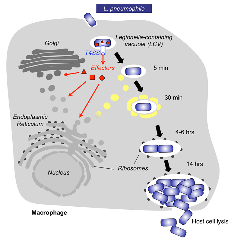
Click image to enlarge.
Intracellular replication cycle of Legionella pneumophila
Upon uptake by a macrophages, L. pneumophila delivers a large number of effector proteins (red) through the Dot/Icm type IV secretion system (T4SS) into the host cytosol. The effectors manipulate signaling and trafficking pathways in order to establish conditions favorable for L. pneumophila growth. Eventually, the host cell is lyzed, and L. pneumophila bacteria infect neighboring cells.
Publications
- Lin YH, Machner MP. Exploitation of the host cell ubiquitin machinery by microbial effector proteins. J Cell Sci 2017 130:1985-1996.
- Lin YH, Doms AG, Cheng E, Kim B, Evans TR, Machner MP. Host cell-catalyzed S-palmitoylation mediates Golgi targeting of the Legionella ubiquitin ligase GobX. J Biol Chem 2015 290:25766-25781.
- Yu X, Decker KB, Baker K, Neunuebel MR, Graves M, Westcott N, Hang H, LaBaer J, Qiu J, Machner MP. Host-pathogen interaction profiling using self-assembling human protein arrays. J Proteome Res 2015 14:1920-1936.
- Machner MP, Storz G. Infection biology: small RNA with a large impact (invited commentary). Nature 2016 529:472-473.
Collaborators
- Aitor Hierro, PhD, CIC bioGUNE Institute, Bilbao, Spain
- Joshua LaBaer, MD, PhD, Virginia G. Piper Center for Personalized Diagnostics, Arizona State University, Tempe, AZ
- Anton Simeonov, PhD, Scientific Director, NCATS, Bethesda, MD
Contact
For more information, email machnerm@mail.nih.gov or visit http://machnerlab.nichd.nih.gov.