The Microscopy and Imaging Core Facility

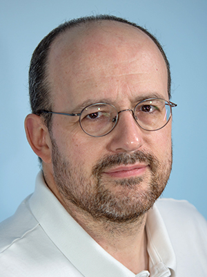
- Vincent Schram, PhD, Biologist
- Louis (Chip) Dye, BS, Research Assistant
- Lynne A. Holtzclaw, BS, Research Assistant
The mission of the NICHD Microscopy and Imaging Core (MIC) is to provide service in three different areas: (1) wide-field and confocal light microscopy, (2) transmission electron microscopy (EM), and (3) sample preparation for light and electron microscopy studies. The Facility is operated as a 'one-stop shop' where investigators can, with a minimum of efforts, go from their scientific question to the final data.
Mode of operation
Located on the ground floor of building 35A, the Facility is accessible 24/7, and users can reserve time on each microscope by using an online calendar (https://next.cirklo.org/nichd). The MIC is available free of charge to all NICHD investigators and, resources allowing, to anyone within the Porter building.
Vincent Schram is the point person for light microscopy and image analysis. The EM branch of the Facility is staffed by Chip Dye, and Lynne Holtzclaw is in charge of sample preparation (histology). Chip Dye and Lynne Holtzclaw report directly to Vincent Schram, who serves as interim director under the management of Chris McBain (NICHD). Tamás Balla (NICHD) acts as scientific advisor for the Facility.
Vincent Schram has a bilateral agreement with Carolyn Smith, who manages the NINDS confocal Facility (LIF), also in the Porter building. Both Facilities freely exchange users, equipment, and support. Although this mode of operation was never codified officially, it is greatly beneficial to the community, as it provides extended support hours, wider expertise, and access to more equipment than each Institute can afford on its own.
Light microscopy
The Facility is well supported by the Office of the Scientific Director. The MIC is equipped with six modern confocal microscopes, each optimized for certain applications: (1) a Zeiss LSM 710 inverted for high-resolution confocal imaging of fixed specimen and live cells; (2) a Zeiss LSM 780 for challenging specimens that require both high resolution and high sensitivity; (3) a Nikon Spinning Disk/Total Internal Reflection Fluorescence (TIRF) hybrid microscope for high-speed confocal imaging or selective recording of membrane-bound events in live cells; (4) a Zeiss LSM 880 2-photon confocal for thick tissues and live animals. In addition, the Core recently received two new confocal systems: (5) a Zeiss 800 optimized for advanced tiling experiments; and (6) a Zeiss 880 Airy, which offers twice the spatial resolution of conventional microscopes without using special dyes or protocols.
Several conventional (wide-field) light microscopes provide imaging modalities such as transmission (visible stains), large-scale tiling of tissue slices, high-speed phase contrast and differential interference contrast (DIC), and large specimens. High-end computer workstations with imaging software (Zeiss Zen, Nikon Element, Bitplane Imaris, SVI Hyugens, and ImageJ) are also available.
After an initial orientation, during which their project is researched by the staff and the best approach is decided upon, users receive hands-on training on the equipment and/or for software best suited to their goals, followed by continuous support when required. Once image acquisition is complete, the staff devise solutions and train users on how to extract usable data from their images. Additional training and support is offered to the community in different ways: (1) on-site assistance and training on equipment owned by individual investigators; (2) an extensive yearly workshop covering light and electron microscopy, image analysis and sample processing; (3) MIC staff volunteer time to teach FAES classes; (4) the Facility organizes frequent on-campus demonstrations of new instruments and software by vendors in a dedicated space. The equipment demonstrations are open to the entire NIH community.
The MIC has a total of 291 registered users in 65 laboratories. At 11,933 instrument hours, overall usage has grown since the last fiscal year, a figure that now includes the animal perfusion station and the JEOL electron microscope. 60% of usage comes from NICHD investigators, most within the Porter building, 20% for training, internal projects, and pilot experiments, and 20% from other Institutes, predominantly NINDS. Usage of each confocal system was uneven, with the Zeiss 710 and 780 being most heavily used. However, it is worth noting that the Zeiss 800 has only been in service since January 2018 and the 880 Airy since April 2018.
Electron microscopy
The electron microscopy branch of the Facility processes specimens from start to finish: fixation, embedding, cutting, ultra-fine sectioning, staining, and imaging on the JEOL 1400 transmission electron microscope. Because of the labor involved, the volume is necessarily smaller than for the light microscopy section, where end users do their own processing. In the past 12 months, Chip Dye processed a total of 67 samples: 53 from NICHD investigators, 2 for MIC internal test projects, and 12 from other Institutes. While this number represents a reduction from last year’s figure, it should be stressed it includes technically challenging and labor-intensive projects for Tamás Balla and Michaela Serpe. The JEOL 1400 electron microscope continues to be available on the calendar for trained users. After the MIC, Joshua Zimmerberg (NICHD) is the major user of that instrument.
Tissue preparation
Lynne Holtzclaw continues to provide sample processing, training, and services to the Facility’s users, both for light and electron microscopy applications. She dedicates a significant amount of time to training users in various techniques, such as rodent perfusion, cryopreservation, cryosectioning, immunofluorescence, and tissue clearing (she trained 21 users during the past 12 months). She processed samples and acquired images for Drs. Balla, Buonanno, Fields, Heuser, Hoffman, Klein, Le Pichon, Loh, Marini, Ozato, Petros, Porter, Sackett, Stojilkovic, and Stopfer (NICHD). She has also provided training and services to Drs. Gordon, Mankodi, Youle (NINDS), Penzo, Plenz, Usdin (NIMH), Chen (NIBIB), and Danner (CC).
A collaborative project with David Klein to characterize pineal cell types for which genes of interest had already been documented by RNA-seq was completed in July 2018. The manuscript is currently being prepared for publication. Lynne Holtzclaw has also joined a collaborative effort with the NINDS laboratory of Richard Youle to study the accumulation of ubiquitinated protein aggregates in brain and liver of a TAX1BP1 knock-out mouse.
Collaborators
- Tamás Balla, PhD, Section on Molecular Signal Transduction, NICHD, Bethesda, MD
- David C. Klein, PhD, Scientist Emeritus, NICHD, Bethesda, MD
- Carolyn L. Smith, PhD, Light Imaging Facility, NINDS, Bethesda, MD
- Richard Youle, PhD, Neurogenetics Branch, NINDS, Bethesda, MD
Contact
For more information, email schramv@mail.nih.gov or visit http://mic.nichd.nih.gov.


