Nervous System Development and Plasticity

- R. Douglas Fields, PhD, Head, Section on Nervous System Development and Plasticity
- Philip Lee, PhD, Staff Scientist
- William Huffman, MA, Technician
- Maxwell Foote, BS, Postbaccalaureate Fellow
- Catherine Carr, PhD, Special Volunteer
- Javier Carvajal Barriga, PhD, Special Volunteer
Healthy development of the brain and cognition in children is central to the mission of the NICHD. Our research is concerned with understanding the molecular and cellular mechanisms by which functional activity in the brain regulates the development of the nervous system during late stages of fetal development and early postnatal life. In addition to synaptic plasticity, we are interested in novel mechanisms of activity-dependent nervous-system plasticity that are particularly relevant to childhood, including the involvement of glia (non-neuronal brain cells). Our work has three main areas of emphasis: myelination and neuron-glia interactions; cellular mechanisms of learning; and gene regulation by neuronal firing. Furthermore, in response to the urgent call for NIH intramural scientists to devote effort to combating the world-wide SARS-Cov2 viral pandemic, our laboratory, in collaboration with others on the NIH campus and in Ecuador, developed a novel nanocellulose material that blocks SARS-Cov2 and HIV viral infection in cell culture.
Traditionally, the field of activity-dependent nervous system development has focused on synapses, and we continue to explore synaptic plasticity. However, our research is also advancing our understanding of how glia sense neural impulse activity and how activity-dependent regulation of glia contributes to development, plasticity, and the cellular mechanisms of learning. A major emphasis of our current research is to understand how myelin (white matter in the brain) is regulated by functional activity. By changing conduction velocity, activity-dependent myelination is a non-synaptic form of plasticity, regulating nervous system function by optimizing the speed and synchrony of information transmission through neural networks. Our studies identified several cellular and molecular mechanisms for activity-dependent myelination, and the findings have important implications for normal brain development, learning, cognition, and psychiatric disorders. Our research shows that myelination of axons by glia (oligodendrocytes in the central nervous system [CNS] and Schwann cells in the peripheral nervous system [PNS]) is regulated by impulse activity, and we identified several molecular mechanisms that control proliferation and differentiation of myelinating glia and myelin formation. Most recently, we determined that myelin thickness can be adjusted through a treadmilling process, which adds and removes layers of myelin from the sheath and alters nodes of Ranvier, where neural impulses are generated along axons. In studies on the visual system, our research shows that this process adjusts conduction velocity and improves functional performance by optimizing spike-time arrival at synapses. This new form of nervous system plasticity and learning is particularly important in childhood development, but it also operates in adulthood.
The discovery of activity-dependent myelin plasticity presents three major questions for this new field of research: 1) Synaptic plasticity is governed by spike timing–dependent plasticity learning rules (STDP), arising from Hebbian theory. What is the equivalent theory underlying myelin plasticity? 2) If myelin plasticity promotes information processing and learning by optimizing synchrony of spike time arrival at synaptic relay points, how can oligodendrocytes ‘know’ that optimal synchrony has been achieved? 3) Do changes in myelin accompany synaptic plasticity that is observed in well established experimental models? This year we obtained experimental and theoretical findings on all three of these major questions.
Learning is perhaps the most important function of childhood, and synaptic plasticity is fundamental to that process. Our research on synaptic plasticity is determining the molecular and cellular mechanisms that convert short-term into long-term memory. Similarly, if functional experiences produce lasting effects on brain development and plasticity, specific genes must be regulated by specific patterns of impulse firing. We are determining how the pattern of neural impulses, i.e., the neural information code, regulates specific genes controlling development and plasticity of neurons and glia.
Figure 1.

Click image to view.
The structure of CNS myelin and nodes of Ranvier
Nervous system plasticity by activity-dependent myelination
The fundamental cellular mechanism of learning, memory, and neural plasticity is synaptic plasticity, in which the strength of synaptic transmission or the number of synapses is modified by experience. Our laboratory has a long-standing interest in synaptic plasticity, but our current interest is to explore new, non-synaptic mechanisms involved in these processes. In particular, we are investigating how changes in neural-impulse transmission velocity contribute to learning and plasticity, and the involvement of glial cells that form the myelin sheath in this new form of plasticity. Our research indicates that, by modifying conduction velocity to optimize the timing of neural impulse arrival at relay points in neural networks, and by influencing the phase and frequency of neural oscillation (brain waves), myelin-forming glia participate in learning, neural plasticity, and nervous system development, in accordance with functional activity and experience.
Myelin, the multilayered membrane of insulation wrapped around nerve fibers (axons) by glial cells, is essential for proper neural impulse transmission and nervous system function. Myelination is a critical part of brain development, but the processes controlling myelination of appropriate axons are not well understood. Myelination begins in the late fetal period and continues throughout childhood and adolescence, but myelination of some brain regions is not complete until an individual's early twenties.
Traditionally, myelin has been viewed in terms of conduction failure after damage (for example in multiple sclerosis), but we are exploring how changes in myelin driven by functional activity affect the timing of neural-impulse arrival at synaptic relay points, which is critical for information processing and synaptic activity. In addition, the frequency, phase, and amplitude-coupling of oscillations in the brain (brainwaves) requires appropriate impulse-conduction velocity, which is influenced by myelination. Many neurological and psychological dysfunctions can develop when optimal neural synchrony of spike-time arrival and neural oscillations are disturbed, as, for example, in schizophrenia, epilepsy, dyslexia, and autism.
Our research shows that neurotransmitters are released not only at synapses but also along axons firing action potentials, to activate receptors on myelinating glia as well as astrocytes and other cells. The recipient cells in turn release growth factors, cytokines, and other molecules that regulate myelination, proliferation, and development of myelinating glia.
Figure 2. Treadmilling model for plasticity of the myelin sheath
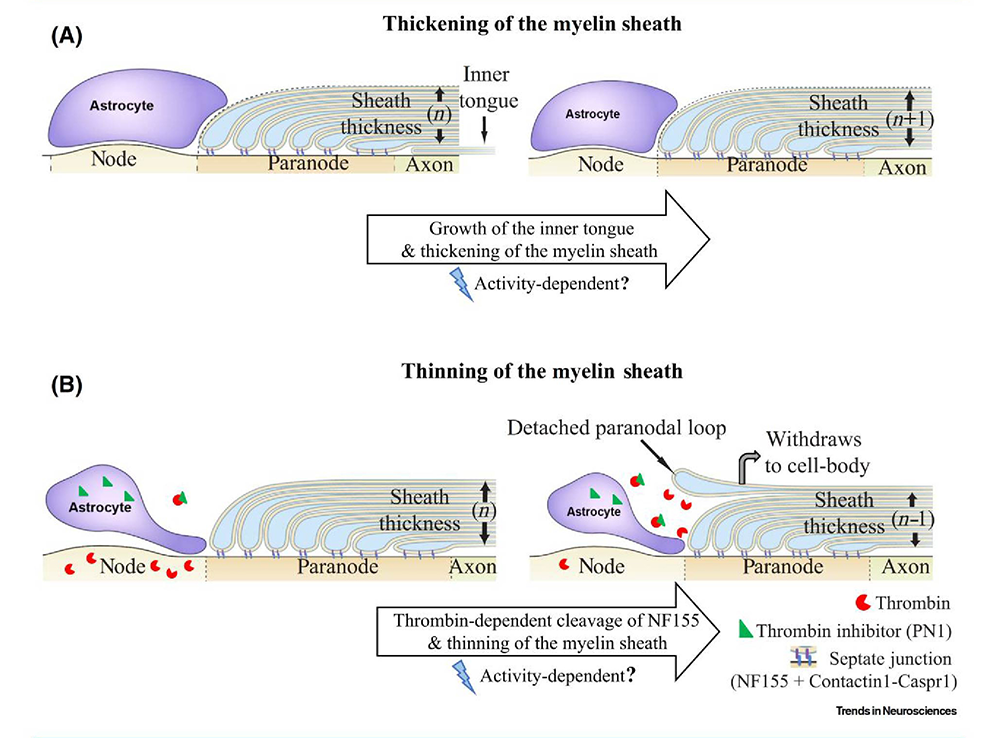
Click image to view.
The speed of neural impulse transmission is altered by changes in myelin structure. The thickness of the myelin sheath in CNS axons is determined by two opposing processes: one (A) that adds additional wraps of myelin to the axon, and the other (B) that removes the outer layer, thereby increasing and decreasing impulse conduction velocity, respectively. New layers of myelin are added beneath the overlaying layers by expansion of the inner tongue of myelin. Myelin is attached to the axon at the paranodal region flanking the node of Ranvier via septate junctions, consisting of neurofascin 155 on myelin interacting with the contactin1-caspr1 complex on the axon. Cleavage of neurofascin 155 by thrombin (red) can break this interaction, resulting in detachment of the outer paranodal loop from the axon, and withdrawal of the outer layer of myelin, which increases nodal gap length and reduces myelin sheath thickness; both effects slow conduction velocity. Perinodal astrocytes at the nodes of Ranvier regulate the process by secreting thrombin inhibitors (green triangle) such as protease nexin1. The treadmilling process helps achieve optimal conduction velocity in individual axons [Fields RD, Dutta DJ Trends Neurosci 2019;42:443].
Induction of myelination by action potentials
In addition to establishing the effects of impulse activity on proliferation and development of myelinating glia, we determined that release of the neurotransmitter glutamate from vesicles along axons triggers the initial events in myelin induction, including stimulating the formation of cholesterol-rich signaling domains between oligodendrocytes and axons and increasing the local synthesis of myelin basic protein, the major protein in the myelin sheath, through Fyn kinase–dependent signaling. We showed that, through this axon-oligodendrocyte signaling mechanism, electrically active axons become preferentially myelinated by a factor of 8 to 1 over electrically inactive axons, thus regulating myelination of axons and neural circuit function according to functional experience. The process would be particularly important in the adolescent brain, where environmental experience during sensitive periods can have long-lasting effects on neural circuit development and behavior. The findings are also relevant to such demyelinating disorders as multiple sclerosis and to re-myelination after axon injury.
Myelin in the PNS is formed by a different type of glial cell: the Schwann cells. Our research is also investigating how neural impulse activity can influence myelination in the PNS, and the results indicate that different mechanisms are responsible from those operating on CNS myelin. These studies involve optogenetics and gene expression analysis, in vivo and in primary cell cultures.
Modification of myelin structure and conduction velocity by astrocytes
Optimal neural-circuit function and synaptic plasticity require the proper impulse transmission speed through all axons to induce spike timing–dependent plasticity and to sustain oscillations at appropriate frequencies. Conduction velocity in myelinated axons depends on the thickness of the myelin sheath and on the morphology of the electrogenic nodes of Ranvier (periodic gaps in the myelin sheath acting as neural transmission relay points along axons). Our research shows that myelination of unmyelinated axons and the thickness of the myelin sheath can be increased in response to neural activity and environmental experience. Prior to our research, myelin structure was believed to be static, and there was no known mechanism that could reduce the thickness of the mature myelin sheath (except in the context of pathology). Our research shows that myelin thickness and nodal gap length are reversibly altered by astrocytes, glial cells that contact nodes of Ranvier, and that this alters the speed of impulse transmission and neural network function. Myelin is attached to the axon by intercellular junctions adjacent to the nodes of Ranvier. We found that one of these cell-adhesion molecules, neurofascin 155, has a binding site for the proteolytic enzyme thrombin, which is secreted by neurons and enters the brain from the vascular system. We found that thrombin-dependent cleavage of neurofascin 155 severs the tether between the axon and myelin, allowing the latter to detach and rendering the myelin sheath thinner. The process is inhibited by vesicular release of thrombin protease inhibitors from perinodal astrocytes. Previously, it was unknown how the myelin sheath could be thinned, and the functions of perinodal astrocytes were not well understood. Our findings uncovered a new form of nervous system plasticity in which myelin structure and conduction velocity are adjusted by astrocytes. The thrombin-dependent cleavage of neurofascin 155 may also have relevance for myelin disruption and repair.
Theoretical advances in myelin plasticity
Spike timing–dependent plasticity (STDP) learning rules govern activity-dependent changes in synaptic strength that underly learning and memory. An equivalent theory guiding myelin plasticity is lacking. Moreover, how oligodendrocytes situated along axons far from synaptic terminals could optimize synchrony of spike time arrival is unknown. In collaboration with our intramural colleagues in NICHD and NIMH, we put forth an oligodendrocyte myelin plasticity (OMP) model that addresses these questions. An oligodendrocyte myelinates multiple axons simultaneously through many slender cell processes extending from an individual cell, which rarely forms myelin on the same axon twice. We propose that this unusual morphology enables oligodendrocytes to sense the arrival of neural impulses at each of its cellular processes and to modify myelin to increase synchrony of action-potential firing among all axons within its domain. The process is extended serially through the chain of oligodendrocytes along the entire length of the axon. Our mathematical simulations show that the process results in greatly increased synchrony of spike time arrival at nerve terminals.
Myelin plasticity accompanying well established models of synaptic plasticity
An implicit assumption in all prior research on synaptic plasticity is that neural-impulse transmission speed is fixed, and not altered by environmental experience. The discovery of myelin plasticity requires a re-examination of that assumption to determine whether or not myelin plasticity accompanies changes in synaptic plasticity. Studies on visual deprivation in kittens provided the fundamental understanding of how environmental experience and differences in the timing of neural impulses from the two eyes arriving on binocular neurons alters synaptic strength and connectivity. Using binocular and monocular visual deprivation in mice, in addition to monocular action-potential inhibition by transfecting a potassium channel into retinal ganglion neurons, we found that changes in myelin are evident in optic nerve and optic tract axons in a manner that is compatible with changes in synaptic strength. Thus, myelin plasticity contributes to, or could even drive, the changes in synaptic strength observed following these different types of visual deprivation.
Regulation of gene expression by action-potential firing patterns
All information in the nervous system is encoded in the temporal pattern of neural impulse firing. Given that long-lasting changes in the nervous system require regulated gene expression, appropriate patterns of neural impulse firing driving neural plasticity must control transcription of specific genes. Little is known about how neural firing patterns regulate gene expression. Our experiments are revealing that intracellular signaling and gene-regulatory networks respond selectively to appropriate temporal patterns of action-potential firing to generate adaptive responses.
To determine how gene expression in neurons and glia is regulated by impulse firing, we stimulate nerve cells to fire impulses in differing patterns by optogenetics and by delivering electrical stimulation through platinum electrodes in specially designed cell-culture dishes. Live-cell calcium imaging shows that temporal aspects of intracellular calcium signaling are particularly important for regulating gene expression according to neural-impulse firing patterns in normal and pathological conditions. After stimulation, we measured mRNA and protein expression by gene microarrays, quantitative RT-PCR (reverse transcriptase–polymerase chain reaction), RNA-seq (RNA sequencing), Western blot, and immunocytochemistry. The results confirm our hypothesis that precise patterns of impulse activity can increase or reduce the expression of specific genes in neurons and glia. Moreover, our research shows that regulation of gene expression in neurons by specific temporal patterns of impulse activity is not a property of special genes; in general, the neuronal transcriptome is highly regulated by the pattern of membrane depolarization, with hundreds of genes differentially regulated by the temporal code of neuronal firing.
We are also pioneering new methods of transcriptional analysis in neurons. The standard approach to analyzing gene expression is to measure the abundance of tens of thousands specific gene transcripts in cells by microarray or RNA-seq, as described above, but this approach fails to capture the unique feature of transcriptional regulation in neurons. In contrast to other cells responding to external signals that may drive cells to a steady-state equilibrium, transcriptional networks in neurons are continually modulated dynamically by temporally varying action-potential firing frequencies and burst patterns, together with synchrony and phase relationships among populations of interconnected neurons. Such activity may not alter the abundance of a gene transcript abundance significantly; nevertheless, the coordinated activity of gene-expression levels within transcriptional networks is being modulated dynamically to modify function.
To address this question, we applied a covariance approach using a Pearson correlation analysis, to determine how pairs of genes in mouse dorsal root ganglion (DRG) neurons are coordinately expressed in response to stimulation producing the same number of action potentials in different temporal patterns. Our analysis of 4,728 distinct gene pairs related to calcium signaling, 435,711 pairs of transcription factors, 820 pairs of voltage-gated ion channels, and 86,862 calcium-signaling genes paired with transcription factors indicated that genes become coordinately activated by distinct action-potential firing patterns. Thus, in addition to regulating the expression level of numerous genes, the temporal pattern of action-potential firing profoundly modulates how genes are networked in functional pathways. We are exploring how chromatin structure is altered by neural impulse firing and how chromatin structure differs between neurons and glia.
Nanocellulose block of SARS-Cov2 and HIV infection
An important world-wide imperative is to develop methods to combat the SARS-Cov2 viral pandemic. In particular, new methods that have broad anti-viral action and are resistant to mutations in viruses that circumvent immunization are especially important. In response to the urgent need to develop methods for combatting Covid-19, our laboratory developed a new method to block infection of cells by SARS-CoV-2. We developed a compound that binds to both the SARS-CoV-2 and the HIV virus and that this prevents cellular infection. Nanocellulose derived from the ivory nut endosperm of a South American palm yields thin nanoparticles (from 1–5 nm width), which provide a high surface area for entrapment of microbes. We determined that the substance is a universal microbe binder, which prevents viral infection of cells in culture. The material can encapsulate the S protein of SARS-CoV-2, as well as whole SARS-CoV-2 and HIV-1 virions, thereby blocking infection of cells. These results were achieved through collaboration with our NIH intramural colleagues and Javier Carvajal Barriga of the Pontificia Universidad Católica del Ecuador, who conducted these experiments as a special volunteer in our laboratory.
Additional Funding
- NIH Intramural Targeted Anti-COVID (ITAC) funding program
Publications
- Pajevic S, Plenz D, Basser PJ, Fields RD. Oligodendrocyte-mediated myelin plasticity. eLIFE 2023 12:e81982.
- Munyeshyaka M, Fields RD. Oligodendroglia are emerging players in several forms of learning and memory. Commun Biol 2022 5(1):1148.
- Santos EN, Huffman WC, Fields RD. Effects of visual deprivation on remodeling of nodes of Ranvier in optic nerve. eNeuro 2022 9(6):ENEURO.0194-22.2022.
- Santos EN, Huffman WC, Fields RD. Recovery of node of Ranvier structure in optic nerve under visual deprivation. Investigative Opthalmology and Visual Science 2023; under review.
- Carvajal-Barriga EJ, Fitzgerald W, Dimitriadis EK, Margolis L, Fields RD. Sulfated endospermic nanocellulose crystals prevent the transmission of SARS-CoV-2 and HIV-1. Sci Rep 2023 13:6959.
Collaborators
- Peter J. Basser, PhD, Section on Quantitative Imaging and Tissue Sciences, NICHD, Bethesda, MD
- Emilios Dimitriadis, PhD, Trans-NIH Shared Resource on Biomedical Engineering and Physical Science, NIBIB, Bethesda, MD
- Wendy Fitzgerald, BS, Section on Intercellular Interactions, NICHD, Bethesda, MD
- Leonid Margolis, PhD, Section on Intercellular Interactions, NICHD, Bethesda, MD
- Sinisa Pajevic, PhD, Division of Computational Bioscience, CIT, NIH, Bethesda, MD
- Dietmar Plenz, PhD, Laboratory of Systems Neuroscience, NINDS, Bethesda, MD
Contact
For more information, email fieldsd@mail.nih.gov.